
글로벌 연구동향
분자영상 및 방사화학


![[Theranostics] Enhanced ASGR2 by microplastic exposure leads to resistance to therapy in gastric cancer](/enewspaper/upimages/1652149569admin.PNG) 2022년 05월호
2022년 05월호
[Theranostics] Enhanced ASGR2 by microplastic exposure leads to resistance to therapy in gastric cancer미세플라스틱에 의하여 유도된 ASGR2 유전자의 위암 치료 저항성 유발KIRAMS / 김현기, 김진수*
- 출처
- Theranostics
- 등재일
- 2022
- 저널이슈번호
- 12(7): 3217-3236. doi: 10.7150/thno.73226
- 내용
-
Abstract
Background: Microplastics (MPs) are a new global environmental threat. Previously, we showed the
biodistribution of MPs using [64Cu] polystyrene (PS) and PET in mice. Here, we aimed to identify whether
PS exposure has malignant effects on the stomach and induces resistance to therapy.
Methods: BALB/c nude mice were fed 1.72 × 10⁴ particles/mL of MP. We investigated PS accumulation
in the stomach using radioisotope-labeled and fluorescent-conjugated PS. Further, we evaluated whether
PS exposure induced cancer stemness and multidrug resistance, and whether it affected tumor
development, tumor growth, and survival rate in vivo using a 4-week PS-exposed NCI-N87 mouse model.
Using RNA-Seq analysis, we analyzed whether PS exposure induced gene expression changes in gastric
tissues of mice.
Results: PET imaging results showed that a single dose of [64Cu]-PS remained for 24 h in the mice
stomach. The 4-week daily repetitive dose of fluorescent conjugated PS was deposited in the gastric
tissues of mice. When PS was exposed, a 2.9-fold increase in migration rate was observed for NCI-N87
cells. Immunocytochemistry results showed decreased E-cadherin and increased N-cadherin expression,
and flow cytometry, qPCR, and western blot analysis indicated a 1.9-fold increase in N-cadherin
expression after PS exposure. Further, PS-induced multidrug resistance to bortezomib, paclitaxel,
gefitinib, lapatinib, and trastuzumab was observed in the NCI-N87 mouse model due to upregulated
CD44 expression. RNA-seq results identified increased asialoglycoprotein receptor 2 (ASGR2)
expression after PS exposure, and ASGR2 knockdown decreased cell proliferation, migration, invasion,
and drug resistance.
Conclusion: We demonstrated that ASGR2 enhanced cancer hallmarks on PS exposure and induced
resistance to chemo- and monoclonal antibody-therapy. Our preclinical findings may provide an incentive
for further epidemiological studies on the role of MP exposure and its association with gastric cancer.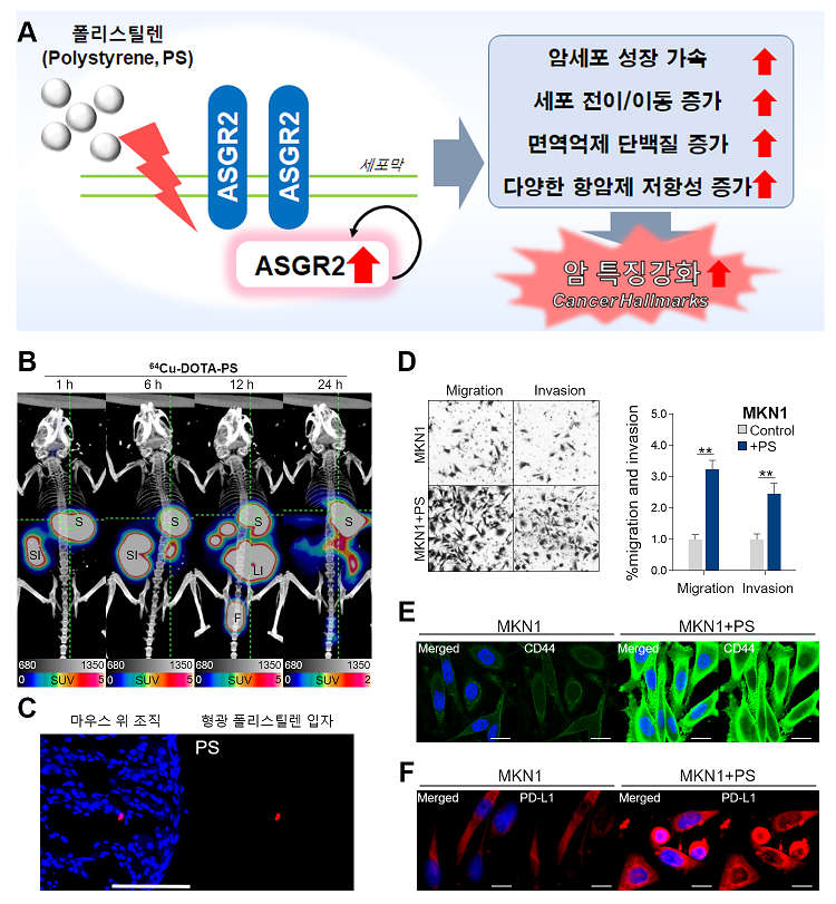
(A) 미세플라스틱 노출에 의한 위암세포의 변화 요약
(B) Cu-64 표지 폴리스틸렌 PET/CT 영상의 대표적 그림
(C) 마우스 위 조직에 형광 폴리스틸렌 입자 확인의 대표적 그림
(D) 폴리스틸렌유발 위암세포주의 Migration / Invasion 증가 확인의 대표적 그림
(E) 폴리스틸렌유발 암 줄기세포 유전자 CD44의 증가 확인의 대표적 그림
(F) 폴리스틸렌유발 면역 회피 유전자 PD-L1 증가 확인의 대표적 그림
Affiliations
Hyeongi Kim1,2, Javeria Zaheer1,3, Eui-Ju Choi2, and Jin Su Kim1,3
1. Division of RI Application, Korea Institute Radiological and Medical Sciences, Seoul 01812, Republic of Korea.
2. Department of Life Sciences, School of Life Sciences and Biotechnology, Korea University, Seoul 02841, Republic of Korea.
3. Radiological and Medico-Oncological Sciences, University of Science and Technology (UST), Seoul 01812, Republic of Korea.
Corresponding author: Jin Su Kim, PhD, Division of RI Application, Korea Institute Radiological and Medical Sciences, 75 Nowon-Gil, Gongneung-Dong,
Nowon-Gu, Seoul 01812, Korea. Radiological and Medico-Oncological Sciences, University of Science and Technology (UST), Seoul 01812, Republic of Korea.
Tel: 82-2-970-1661, Fax: 82-2-970-2416, E-mail: kjs@kirams.re.kr.
- 연구소개
- 지난해부터 방사성 동위원소 표지-미세플라스틱을 이용한 체내 흡수 경로를 확인하고, 자폐스펙트럼 장애 유발 등 미세플라스틱의 연구를 이어오고 있습니다, 이번 연구는 한국인에게 흔한 위암에 주목하고 미세플라스틱과의 상관관계를 찾아보고자 진행하였습니다. 각종 일회용품 등에 쓰이는 플라스틱 종류 중 하나인 폴리스틸렌(직경 10마이크로미터 크기)을 5종류의 인간유례 위암 세포주에서 폴리스틸렌과 4주간 함께 배양하여 암의 주요 특징(Hallmarks of Cancer) 변화를 확인했습니다. 폴리스틸렌 미세플라스틱에 4주간 반복 노출된 위암 세포는 노출되지 않은 위암 세포에 비해 최대 74% 더 빠르게 자랐고, 전이는 최대 3.2∼11배 많았으며, 약물저항성 및 치료 저항성을 유발할 수 있는 암 줄기세포 유전자 CD44는 최대 3.4배 증가했고, 암세포가 면역시스템을 피하기 위해 만들어내는 면역억제 단백질 PD-L1(CD274)의 발현은 최대 4.2배 증가를 확인하였습니다. 또한, 4주간 폴리스틸렌에 노출된 위암 세포들은 미세플라스틱 노출로 인해 증가한 CD44 단백질 및 ASGR2 단백질을 통해 다양한 화학요법 및 항체 치료제 내성을 유발하는 것을 확인하였습니다. 이와 함께 폴리스틸렌을 먹인 실험용 쥐의 위 조직에서 폴리스틸렌유발 특이 유전자들을 RNAseq 방법으로 확인하였고, 그 중 세포막 단백질 아시알로당 단백질 수용체2(Asialoglycoprotein receptor 2, ASGR2) 에 특이적 증가를 확인하였습니다. 폴리스틸렌 미세플라스틱에 4주간 반복 노출된 위암 세포에서 ASGR2의 증가를 교차 검증 하였고, ASGR2 siRNA를 이용하여, 폴리스틸렌 증가 ASGR2를 내려주면, 악성 유전자 변이, 전이 촉진, 성장가속 및 항암제 저항성을 막을 수 있는 것을 확인하였습니다.
- 덧글달기


