글로벌 연구동향
방사선생물학
![[Int J Radiat Oncol Biol Phys.] Real-time Tumor Oxygenation Changes After Single High-dose Radiation Therapy in Orthotopic and Subcutaneous Lung Cancer in Mice: Clinical Implication for Stereotactic Ablative Radiation Therapy Schedule Optimization.](/enewspaper/upimages/admin_20160810111740_R.jpg) 2016년 08월호
2016년 08월호
[Int J Radiat Oncol Biol Phys.] Real-time Tumor Oxygenation Changes After Single High-dose Radiation Therapy in Orthotopic and Subcutaneous Lung Cancer in Mice: Clinical Implication for Stereotactic Ablative Radiation Therapy Schedule Optimization.서울의대, 포항공대/ 송창훈, 홍범주, 김학재*, 안지완*
- 출처
- Int J Radiat Oncol Biol Phys.
- 등재일
- 2016 Jul 1
- 저널이슈번호
- 95(3):1022-31. doi: 10.1016/j.ijrobp.2016.01.064. Epub 2016 Feb 13.
- 내용

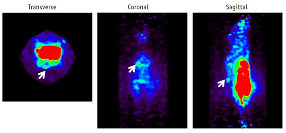
마우스 종양에 F-MISO 가 섭취된 PET 영상
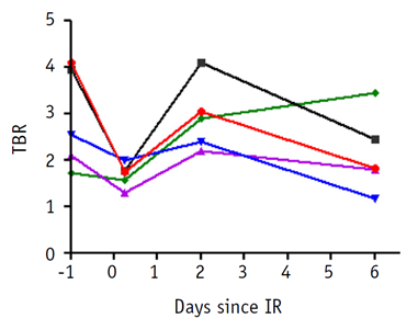
방사선조사 전후 종양의 재산소화의 변화 분석 결과Abstract
PURPOSE:
To investigate the serial changes of tumor hypoxia in response to single high-dose irradiation by various clinical and preclinical methods to propose an optimal fractionation schedule for stereotactic ablative radiation therapy.
METHODS AND MATERIALS:
Syngeneic Lewis lung carcinomas were grown either orthotopically or subcutaneously in C57BL/6 mice and irradiated with a single dose of 15 Gy to mimic stereotactic ablative radiation therapy used in the clinic. Serial [(18)F]-misonidazole (F-MISO) positron emission tomography (PET) imaging, pimonidazole fluorescence-activated cell sorting analyses, hypoxia-responsive element-driven bioluminescence, and Hoechst 33342 perfusion were performed before irradiation (day -1), at 6 hours (day 0), and 2 (day 2) and 6 (day 6) days after irradiation for both subcutaneous and orthotopic lung tumors. For F-MISO, the tumor/brain ratio was analyzed.
RESULTS:
Hypoxic signals were too low to quantitate for orthotopic tumors using F-MISO PET or hypoxia-responsive element-driven bioluminescence imaging. In subcutaneous tumors, the maximum tumor/brain ratio was 2.87 ± 0.483 at day -1, 1.67 ± 0.116 at day 0, 2.92 ± 0.334 at day 2, and 2.13 ± 0.385 at day 6, indicating that tumor hypoxia was decreased immediately after irradiation and had returned to the pretreatment levels at day 2, followed by a slight decrease by day 6 after radiation. Pimonidazole analysis also revealed similar patterns. Using Hoechst 33342 vascular perfusion dye, CD31, and cleaved caspase 3 co-immunostaining, we found a rapid and transient vascular collapse, which might have resulted in poor intratumor perfusion of F-MISO PET tracer or pimonidazole delivered at day 0, leading to decreased hypoxic signals at day 0 by PET or pimonidazole analyses.
CONCLUSIONS:
We found tumor hypoxia levels decreased immediately after delivery of a single dose of 15 Gy and had returned to the pretreatment levels 2 days after irradiation and had decreased slightly by day 6. Our results indicate that single high-dose irradiation can produce a rapid, but reversible, vascular collapse in tumors.
Author information
Song C1, Hong BJ2, Bok S2, Lee CJ2, Kim YE2, Jeon SR3, Wu HG4, Lee YS5, Cheon GJ6, Paeng JC6, Carlson DJ7, Kim HJ8, Ahn GO9.
1Department of Radiation Oncology, Seoul National University College of Medicine, Seoul, Korea.
2Division of Integrative Biosciences and Biotechnology, Pohang University of Science and Technology, Pohang, Gyeongbuk, Korea.
3Department of Radiation Oncology, Seoul National University College of Medicine, Seoul, Korea; Cancer Research Institute, Seoul National University College of Medicine, Seoul, Korea.
4Department of Radiation Oncology, Seoul National University College of Medicine, Seoul, Korea; Cancer Research Institute, Seoul National University College of Medicine, Seoul, Korea; Institute of Radiation Medicine, Medical Research Center, Seoul National University College of Medicine, Seoul, Korea.
5Department of Nuclear Medicine, Seoul National University College of Medicine, Seoul, Korea; Department of Molecular Medicine and Biopharmaceutical Sciences, Seoul National University College of Medicine, Seoul, Korea.
6Cancer Research Institute, Seoul National University College of Medicine, Seoul, Korea; Institute of Radiation Medicine, Medical Research Center, Seoul National University College of Medicine, Seoul, Korea; Department of Nuclear Medicine, Seoul National University College of Medicine, Seoul, Korea.
7Department of Therapeutic Radiology, Yale University School of Medicine, New Haven, Connecticut.
8Department of Radiation Oncology, Seoul National University College of Medicine, Seoul, Korea; Cancer Research Institute, Seoul National University College of Medicine, Seoul, Korea; Institute of Radiation Medicine, Medical Research Center, Seoul National University College of Medicine, Seoul, Korea. Electronic address: khjae@snu.ac.kr.
9Division of Integrative Biosciences and Biotechnology, Pohang University of Science and Technology, Pohang, Gyeongbuk, Korea. Electronic address: goneahn@postech.ac.kr.
- 연구소개
- 방사선치료의 중요 기법 중 하나인 체부정위방사선치료에서 최적의 치료 스케줄을 임상에 적용하기 위한 목적으로 시행한 전임상 연구입니다. 종양의 고선량 방사선조사 후 재산소화가 되는 시점을 보고자 F-MISO PET을 시행하여 1) 고선량 방사선조사 직후에는 종양 내 혈관이 일시적으로 수축되는 현상과 2) 방사선조사 2일 이후에 지속적으로 재산소화가 일어남을 확인하였습니다. 본 연구를 바탕으로 고선량 방사선조사 후 재산소화 시점에 대한 추가적인 연구가 필요할 것으로 생각됩니다.
- 덧글달기


