


글로벌 연구동향
의학물리학


![[Med Phys .] A feasibility study on deep-neural-network-based dose-neutral dual-energy digital breast tomosynthesis](/enewspaper/upimages/1683594396admin.JPG) [Med Phys .] A feasibility study on deep-neural-network-based dose-neutral dual-energy digital breast tomosynthesis
[Med Phys .] A feasibility study on deep-neural-network-based dose-neutral dual-energy digital breast tomosynthesis
심층신경망 기반 이중에너지 디지털 유방 단층영상 합성 연구KAIST / 김형석, 이호연, 조승룡*
- 출처
- Med Phys .
- 등재일
- 2023 Feb
- 저널이슈번호
- 50(2):791-807. doi: 10.1002/mp.16071. Epub 2022 Nov 3
- 내용
Abstract
Background: Diagnostic performance based on x-ray breast imaging is subject to breast density. Although digital breast tomosynthesis (DBT) is reported to outperform conventional mammography in denser breasts, mass detection and malignancy characterization are often considered challenging yet.Purpose: As an improved diagnostic solution to the dense breast cases, we propose a dual-energy DBT imaging technique that enables breast compositional imaging at comparable scanning time and patient dose compared to the conventional single-energy DBT.
Methods: The proposed dual-energy DBT acquires projection data by alternating two different energy spectra. Then, we synthesize unmeasured projection data using a deep neural network that exploits the measured projection data and adjacent projection data obtained under the other x-ray energy spectrum. For material decomposition, we estimate partial path lengths of an x-ray through water, lipid, and protein from the measured and the synthesized projection data with the object thickness information. After material decomposition in the projection domain, we reconstruct material-selective DBT images. The deep neural network is trained with the numerical breast phantoms. A pork meat phantom is scanned with a prototype dual-energy DBT system to demonstrate the feasibility of the proposed imaging method.
Results: The developed deep neural network successfully synthesized missing projections. Material-selective images reconstructed from the synthesized data present comparable compositional contrast of the cancerous masses compared with those from the fully measured data.
Conclusions: The proposed dual-energy DBT scheme is expected to substantially contribute to enhancing mass malignancy detection accuracy particularly in dense breasts.
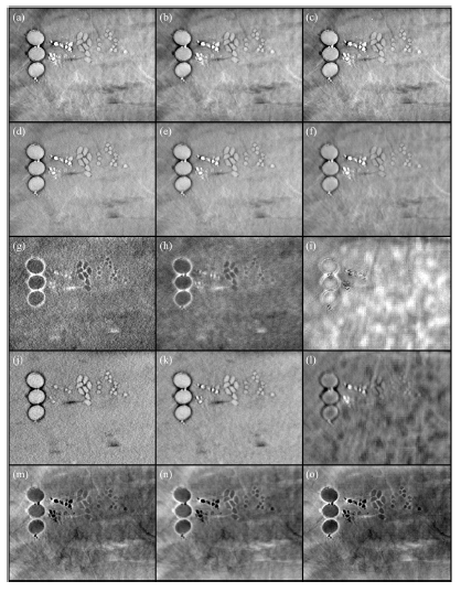
(위쪽 row부터: Low-energy DBT, High-energy DBT, Water-based, Protein-based, and Lipid-based 영상, 왼쪽 column부터: Double dose 조건의 reference 이중에너지 DBT 결과, 제안된 방법으로 얻은 결과, 그리고 한쪽 에너지 정보로만 합성한 네트워크 결과)
본 영상은 돼지고기에 다양한 모사 물질 (콩, 곡물, 석회 등)을 넣어 만든 팬텀을 촬영한 것으로 제안된 방식에서 기준 영상과 유사한 영상 품질 및 물질 분별력을 갖춘 것을 보여주고 있음.
Affiliations
Hyeongseok Kim 1, Hoyeon Lee 2, Seoyoung Lee 3, Young-Wook Choi 4, Young Jin Choi 4, Kee Hyun Kim 4, Wontaek Seo 5, Choul Woo Shin 5, Seungryong Cho 1 3 6 7
1KAIST Institute for Artificial Intelligence, Korea Advanced Institute of Science and Technology (KAIST), Daejeon, South Korea.
2Department of Radiation Oncology, Massachusetts General Hospital and Harvard Medical School, Boston, Massachusetts, USA.
3Department of Nuclear and Quantum Engineering, Korea Advanced Institute of Science and Technology (KAIST), Daejeon, South Korea.
4Korea Electrotechnology Research Institute (KERI), Ansan, South Korea.
5DRTECH, Seongnam, South Korea.
6KAIST Institute for Health Science and Technology, Korea Advanced Institute of Science and Technology (KAIST), Daejeon, South Korea.
7KAIST Institute for IT Convergence, Korea Advanced Institute of Science and Technology (KAIST), Daejeon, South Korea.
- 키워드
- deep learning; digital breast tomosynthesis; dual-energy x-ray imaging; image reconstruction; material decomposition.
- 연구소개
- 여성 유방암 검진에 활용되고 있는 DBT (Digital breast tomosynthesis) 영상 촬영 기법에 있어서, 기저 물질 기반의 물질 분별 mapping을 제공하여 검진의 정확도를 높이는데 기여하고자 개발한 방법을 소개하고 있습니다. X선 이중에너지 스캔 방식은 물질 분별을 위해 두루 사용되고 있는데, 현재 가용한 DBT 장비에서 이중에너지 스캔을 구현하기 위해서는 X선 관전압을 바꾸어 두 번 촬영하는 방식을 생각해 볼 수 있습니다. 유방 촬영술에서 X선 선량이 배가되는 것은 바람직하지 않으므로, 본 연구에서는 dose-neutral 한 방식 구현을 위해 DBT 촬영 시에 이웃하는 프로젝션마다 관전압을 스위칭하여 전체 프로젝션 수는 변하지 않는 방식을 제안했습니다. 이로 인해 각 에너지 대역의 DBT 영상 구현을 위해 프로젝션 수가 부족해지는 문제가 발생하고, 이를 극복하기 위해 딥러닝 영상 합성 기술을 이용하여 프로젝션 수를 배로 증가시키는데 성공했으며, 결과적으로 얻어진 물질 분별 영상의 정확도 평가를 통해 제안 방법이 임상에서도 유용한 기술로 활용될 수 있을 가능성을 입증하였습니다.
- 덧글달기


![]()
- COPYRIGHT(C) 2015 한국원자력의학원 전략기획팀 All rights Reserved.
- 문의 : rmwebzine@kirams.re.kr 발행처 : 한국원자력의학원 전략기획팀
- 우) 01812 서울시 노원구 노원로 75 한국원자력의학원 전략기획팀