글로벌 연구동향
의학물리학
![[Med Phys] Detection of masses in digital breast tomosynthesis using complementary information of simulated projection.](/enewspaper/upimages/admin_20160115091112_R.jpg) 2016년 01월호
2016년 01월호
[Med Phys] Detection of masses in digital breast tomosynthesis using complementary information of simulated projection.KAIST / 김성태, 노용만*
- 출처
- Med Phys
- 등재일
- 2015 Dec
- 저널이슈번호
- 42(12):7043. doi: 10.1118/1.4935533.
- 내용

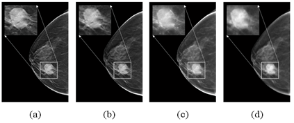
그림 1. Circumscribed margin을 갖는 병변의 합성영상 화질 비교. (a) 제안하는 합성영상, (b) DBT 단면영상의 central slice, (c) maximum intensity projection, (d) average projection. 기존 합성영상 (c, d)보다 제안하는 합성영상 (a)에서 병변이 더 뚜렷하게 나타남을 보임.

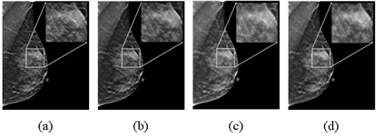
그림 2. Ill-defined margin을 갖는 병변의 합성영상 화질 비교. (a) 제안하는 합성영상, (b) DBT 단면영상의 central slice, (c) maximum intensity projection, (d) average projection. 기존 합성영상 (c, d)보다 제안하는 합성영상 (a)에서 병변이 더 뚜렷하게 나타남을 보임.

그림 3. 제안 방법에서 병변 검출에 활용한 영상에 따른 case-based FROC 성능 비교. 단면영상과 합성영상 모두를 사용할 때 (combined approach), 병변 검출 성능이 향상됨을 보임.
Abstract
PURPOSE:
The purpose of this study is to develop a computer-aided detection system that combines the detection results in 3D digital breast tomosynthesis (DBT) volume and 2D simulated projection (synthesized image which is not provided by the vendor but generated from DBT volume in this study) to improve the accuracy of mass detection in DBT.
METHODS:
The 3D DBT volume has a problem of blurring in the out-of-focus plane because it is reconstructed from a limited number of projection view images acquired over a limited angular range. To solve the problem, the simulated projection is generated by measuring the blurriness of voxels in the DBT volume and adopting conspicuity voxels. A contour-based detection algorithm is applied to detecting masses in the simulated projection. The DBT volume is analyzed by using an unsupervised mass detection algorithm, which results in mass candidates in the DBT volume. The mass likelihood scores estimated for mass candidates on the DBT volume and the simulated projection are merged in a probabilistic manner through a Bayesian network model to differentiate masses and false positives (FPs). Experiments were conducted on a clinical data set of 320 DBT volumes. In 90 volumes, at least one biopsy-proven malignant mass was presented. The longest diameter of masses ranged from 7.0 to 56.4 mm (mean = 25.4 mm). The sizes of masses in the data set were relatively large compared to the sizes of the masses reported in other detection studies. Three image quality measurements (overall sharpness, sharpness of mass boundary, and contrast) were used to evaluate the image quality of the simulated projection compared to the DBT central slice where the mass was most conspicuous and other projection methods (maximum intensity projection and average projection). A free-response receiver operating characteristic (FROC) analysis was adopted for evaluating the accuracy of mass detection in the DBT volume, the simulated projection, and the combined approach. A jackknife FROC analysis (JAFROC) was used to estimate the statistical significance of the difference between two FROC curves.
RESULTS:
The overall sharpness and the sharpness of mass boundary in the simulated projection are higher than those in the DBT central slice and other projection methods. The contrast of the simulated projection is lower than the DBT central slice. The mass detection in the DBT volume achieved region-based sensitivities of 80% and 85% with 1.75 and 2.11 FPs per DBT volume. The proposed combined mass detection approach achieved same sensitivities with reduced FPs of 1.33 and 1.93 per DBT volume. The difference of the FROC curves between the combined approach and the mass detection in the DBT volume was statistically significant (p < 0.01) by JAFROC analysis.
CONCLUSIONS:
This study indicates that the combined approach that merges the detection results in the DBT volume and the simulated projection is a promising approach to improve the accuracy of mass detection in DBT.
Author information
Kim ST1, Kim DH1, Ro YM1.
1Department of Electrical Engineering, KAIST, 291 Daehak-ro, Yuseong-Gu, Daejeon 305-701, Republic of Korea.
- 연구소개
- Digital breast tomosynthesis (DBT)는 기존 2차원 mammography 의 조직중첩문제를 해결하기 위해 3차원 단면영상(reconstructed volume)을 얻는 새로운 유방 영상 촬영술입니다. 최근 DBT와 기존 mammography를 같이 사용할 경우 유방암 진단 정확도가 향상된다는 연구가 발표되고 있습니다. 하지만 DBT와 mammography를 같이 사용할 경우 환자의 방사선 노출이 증가한다는 한계가 있어 mammography를 대신 DBT 영상으로부터 병변진단에 유용한 2차원 영상을 합성하는 연구가 주목을 받고 있습니다. 본 연구에서는 DBT 단면영상을 분석하여 병변이 뚜렷한 부분을 투영시켜 2차원 합성영상 (simulated projection)을 획득하는 방법을 제안하고 3차원 단면영상과 2차원 합성영상의 정보를 융합한 병변 (mass) 검출 방법을 제안했습니다. 실험을 통해 기존 합성영상과 비교하여 제안하는 합성영상에서 병변이 더 뚜렷하게 나타나는 것을 (그림 1, 2) 보였습니다. 또한 단면영상과 합성영상의 상보적인 특징으로부터 단면영상과 합성영상의 검출 결과의 융합이 유방암 검출에 효과적임을 (그림 3) 보였습니다. 본 연구는 합성영상 생성, 다중 영상 융합에 관심 있는 연구자들에게 도움이 될 것이라 기대합니다.
- 덧글달기


