글로벌 연구동향
의학물리학
 Fully iterative scatter corrected digital breast tomosynthesis using GPU-based fast Monte Carlo simulation and composition ratio update
KAIST / 김경상, 이태원, 예종철*
- 출처
- Med Phys
- 등재일
- 2015 Sep
- 저널이슈번호
- 42(9):5342. doi: 10.1118/1.4928139.
- 내용

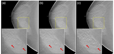
(a) 산란 보정 전 재구성 영상, (b) Convolution기반의 산란 보정 후 재구성 영상, 그리고 (c) 제안하는 방법을 이용한 산란 보정 후 재구성 영상AbstractPURPOSE:
In digital breast tomosynthesis (DBT), scatter correction is highly desirable, as it improves image quality at low doses. Because the DBT detector panel is typically stationary during the source rotation, antiscatter grids are not generally compatible with DBT; thus, a software-based scatter correction is required. This work proposes a fully iterative scatter correction method that uses a novel fast Monte Carlo simulation (MCS) with a tissue-composition ratio estimation technique for DBT imaging.
METHODS:
To apply MCS to scatter estimation, the material composition in each voxel should be known. To overcome the lack of prior accurate knowledge of tissue composition for DBT, a tissue-composition ratio is estimated based on the observation that the breast tissues are principally composed of adipose and glandular tissues. Using this approximation, the composition ratio can be estimated from the reconstructed attenuation coefficients, and the scatter distribution can then be estimated by MCS using the composition ratio. The scatter estimation and image reconstruction procedures can be performed iteratively until an acceptable accuracy is achieved. For practical use, (i) the authors have implemented a fast MCS using a graphics processing unit (GPU), (ii) the MCS is simplified to transport only x-rays in the energy range of 10-50 keV, modeling Rayleigh and Compton scattering and the photoelectric effect using the tissue-composition ratio of adipose and glandular tissues, and (iii) downsampling is used because the scatter distribution varies rather smoothly.
RESULTS:
The authors have demonstrated that the proposed method can accurately estimate the scatter distribution, and that the contrast-to-noise ratio of the final reconstructed image is significantly improved. The authors validated the performance of the MCS by changing the tissue thickness, composition ratio, and x-ray energy. The authors confirmed that the tissue-composition ratio estimation was quite accurate under a variety of conditions. Our GPU-based fast MCS implementation took approximately 3 s to generate each angular projection for a 6 cm thick breast, which is believed to make this process acceptable for clinical applications. In addition, the clinical preferences of three radiologists were evaluated; the preference for the proposed method compared to the preference for the convolution-based method was statistically meaningful (p < 0.05, McNemar test).
CONCLUSIONS:
The proposed fully iterative scatter correction method and the GPU-based fast MCS using tissue-composition ratio estimation successfully improved the image quality within a reasonable computational time, which may potentially increase the clinical utility of DBT.
Author information
Kim K1, Lee T2, Seong Y3, Lee J3, Jang KE3, Choi J4, Choi YW4, Kim HH5, Shin HJ5, Cha JH5, Cho S2, Ye JC1.
1Bio Imaging and Signal Processing Laboratory, Department of Bio and Brain Engineering, KAIST 291, Daehak-ro, Yuseong-gu, Daejeon 34141, South Korea.
2Medical Imaging and Radiotherapeutics Laboratory, Department of Nuclear and Quantum Engineering, KAIST 291, Daehak-ro, Yuseong-gu, Daejeon 34141, South Korea.
3Samsung Advanced Institute of Technology, Samsung Electronics, 130, Samsung-ro, Yeongtong-gu, Suwon-si, Gyeonggi-do, 443-803, South Korea.
4Korea Electrotechnology Research Institute (KERI), 111, Hanggaul-ro, Sangnok-gu, Ansan-si, Gyeonggi-do, 426-170, South Korea.
5Department of Radiology and Research Institute of Radiology, Asan Medical Center, University of Ulsan College of Medicine, 88 Olympic-ro, 43-gil, Songpa-gu, Seoul, 138-736, South Korea.
- 연구소개
- 본 연구는 3차원 디지털유방단층합성촬영(Digital Breast Tomosynthesis, DBT) 영상 재구성을 위한 X-ray 산란 추정 및 보정 알고리즘에 관한 내용이다. DBT의 경우 일반적으로 디텍터는 고정되어있고 X-ray 소스가 한정적인 각도(ex. -21°~21°)만큼 회전하여 영상을 획득하게 된다. 이 때 기존의 컴퓨터단층촬영(CT)과 달리 X-ray 소스와 디텍터 사이의 기하학적인 구조가 각도에 따라 변화하기 때문에 Anti-scatter grid와 같은 하드웨어적인 방법으로 산란을 보정하기 어렵다. 이 문제를 해결하기 위해 몬테카를로 시뮬레이션을 통해 X-ray 광자를 모델링 하여 산란을 추정하고 영상 재구성에 적용하여 산란을 보정하는 방법을 제안하였다. 몬테카를로 방법은 기존의 Convolution기반의 알고리즘들에 비해 파라미터 세팅이 없다는 가장 큰 장점이 있지만 물체의 구성 물질에 대한 정보를 알아야 연산 할 수 있다. 본 논문에서는 유방이 유선조직(glandular)과 지방(adipose)로만 이루어져있다고 가정하였고 유선 조직과 지방의 비율로 나타낸 구성 물질 비율(Composition ratio)을 Voxel별로 추정하여 계산하는 독창적인 방법을 제안하였다. 이를 바탕으로 영상 재구성과 영상에서부터 구성물질비율 계산 그리고 몬테카를로 시뮬레이션을 통한 산란 추정 과정을 2~3번 반복하여 최종 수렴된 산란 값 및 재구성 영상을 획득할 수 있었다. 검증을 위해 Blind test를 하였고 기존의 Convolution 방법에 비해 우수함을 확인하였다. 마지막으로 그래픽연산장치를 이용한 병렬처리 기법으로 알고리즘을 고속화하였고 각도 별로 3초 내외로 산란 값을 계산할 수 있었다. 따라서 본 논문은 DBT 영상 재구성에서 몬테카를로 방법을 이용한 산란 보정을 임상에 적용 가능한 수준으로 구현하였다는 것에 큰 의미가 있다.
- 덧글달기


