글로벌 연구동향
의학물리학
 2015년 09월호
Generation of hybrid sinograms for the recovery of kV-CT images with metal artifacts for helical tomotherapy부산의대/ 전호상, 김호경*
- 출처
- Med Phys
- 등재일
- 2015 Aug
- 저널이슈번호
- 42(8):4654
- 내용


[Abstract]
PURPOSE:
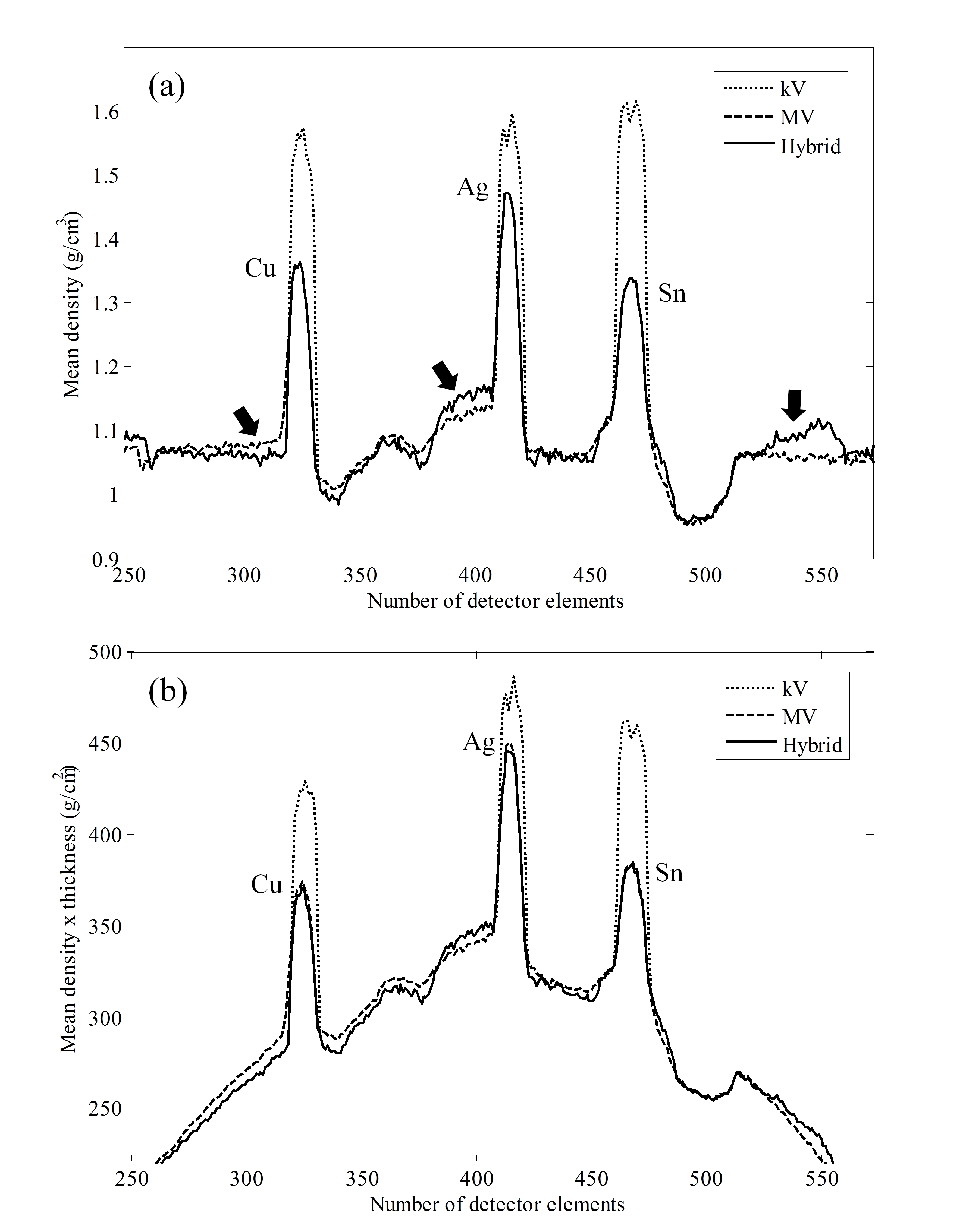
The overall goal of this study is to restore kilovoltage computed tomography (kV-CT) images which are disfigured by patients' metal prostheses. By generating a hybrid sinogram that is a combination of kV and megavoltage (MV) projection data, the authors suggest a novel metal artifact-reduction (MAR) method that retains the image quality to match that of kV-CT and simultaneously restores the information of metal prostheses lost due to photon starvation.
METHODS:
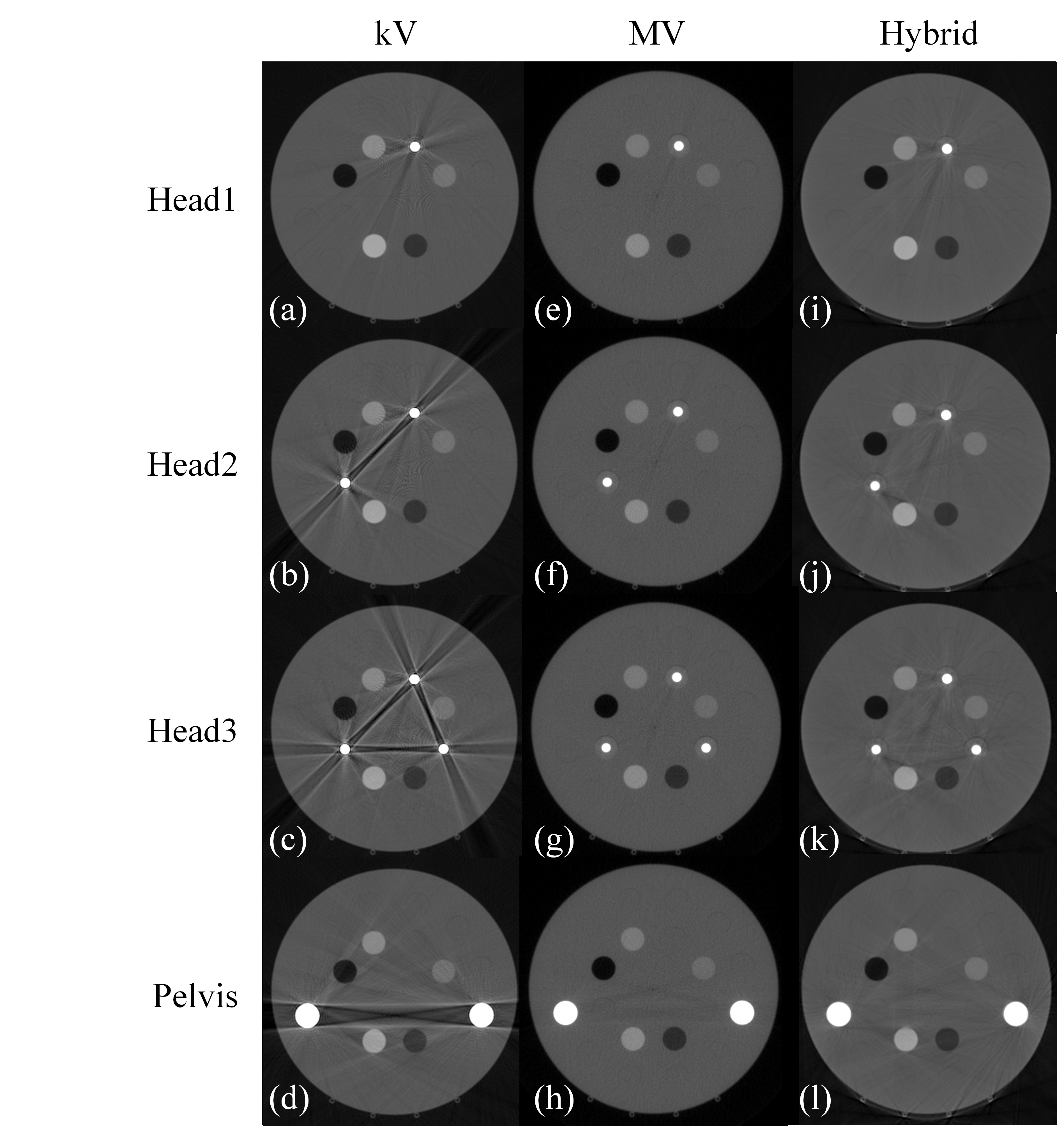
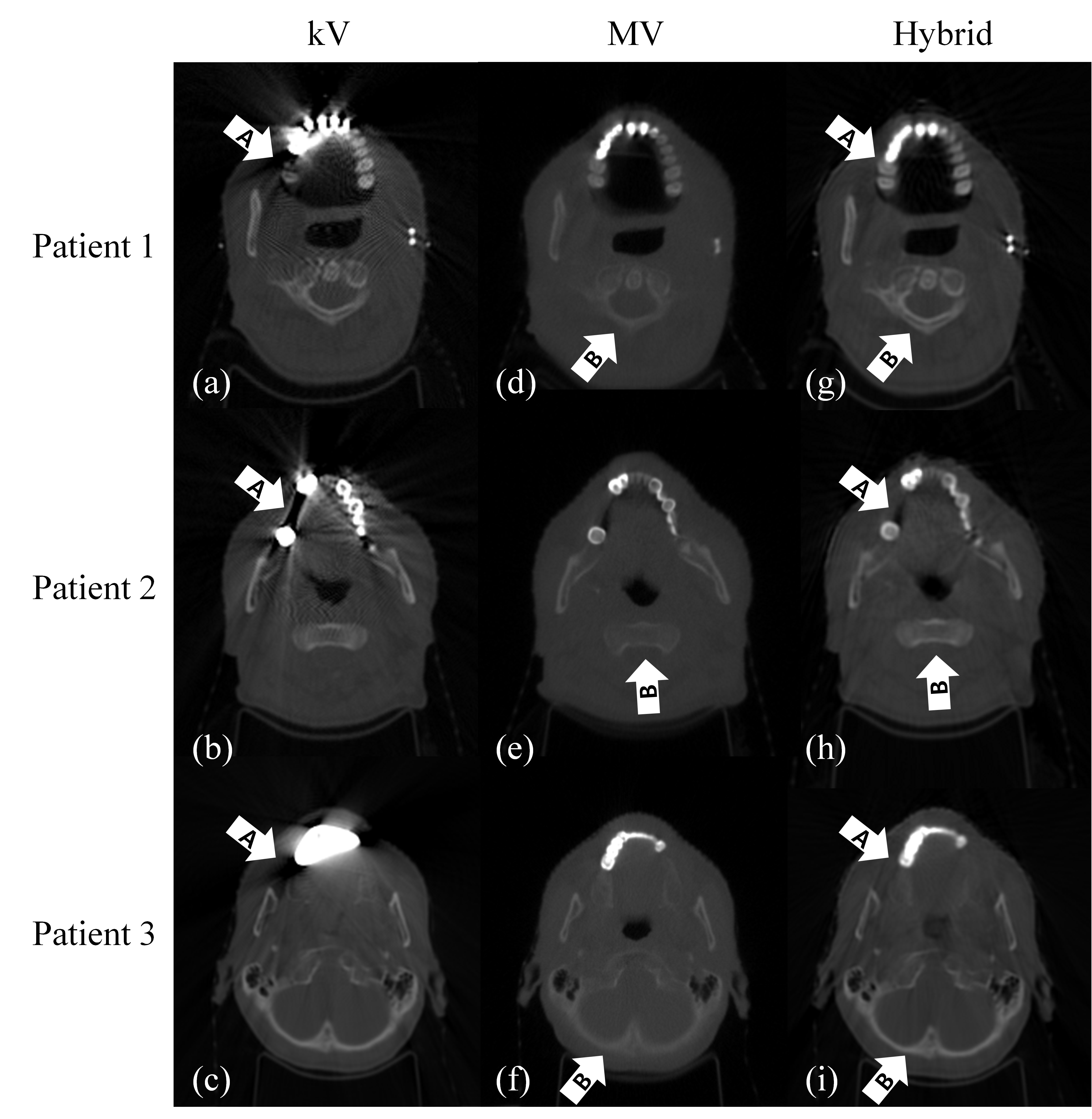
CT projection data contain information about attenuation coefficients and the total length of the attenuation. By normalizing raw kV projections with their own total lengths of attenuation, mean attenuation projections were obtained. In the same manner, mean density projections of MV-CT were obtained by the normalization of MV projections resulting from the forward projection of density-calibrated MV-CT images with the geometric parameters of the kV-CT device. To generate the hybrid sinogram, metal-affected signals of the kV sinogram were identified and replaced by the corresponding signals of the MV sinogram following a density calibration step with kV data. Filtered backprojection was implemented to reconstruct the hybrid CT image. To validate the authors' approach, they simulated four different scenarios for three heads and one pelvis using metallic rod inserts within a cylindrical phantom. Five inserts describing human body elements were also included in the phantom. The authors compared the image qualities among the kV, MV, and hybrid CT images by measuring the contrast-to-noise ratio (CNR), the signal-to-noise ratio (SNR), the densities of all inserts, and the spatial resolution. In addition, the MAR performance was compared among three existing MAR methods and the authors' hybrid method. Finally, for clinical trials, the authors produced hybrid images of three patients having dental metal prostheses to compare their MAR performances with those of the kV, MV, and three existing MAR methods.
RESULTS:
The authors compared the image quality and MAR performance of the hybrid method with those of other imaging modalities and the three MAR methods, respectively. The total measured mean of the CNR (SNR) values for the nonmetal inserts was determined to be 14.3 (35.3), 15.3 (37.8), and 25.5 (64.3) for the kV, MV, and hybrid images, respectively, and the spatial resolutions of the hybrid images were similar to those of the kV images. The measured densities of the metal and nonmetal inserts in the hybrid images were in good agreement with their true densities, except in cases of extremely low densities, such as air and lung. Using the hybrid method, major streak artifacts were suitably removed and no secondary artifacts were introduced in the resultant image. In clinical trials, the authors verified that kV and MV projections were successfully combined and turned into the resultant hybrid image with high image contrast, accurate metal information, and few metal artifacts. The hybrid method also outperformed the three existing MAR methods with regard to metal information restoration and secondary artifact prevention.
CONCLUSIONS:
The authors have shown that the hybrid method can restore the overall image quality of kV-CT disfigured by severe metal artifacts and restore the information of metal prostheses lost due to photon starvation. The hybrid images may allow for the improved delineation of structures of interest and accurate dose calculations for radiation treatment planning for patients with metal prostheses.
[Author information]
Jeon H1, Park D2, Youn H3, Nam J4, Lee J4, Kim W2, Ki Y2, Kim YH2, Lee JH2, Kim D2, Kim HK5.
1Department of Radiation Oncology and Research Institute for Convergence of Biomedical Science and Technology, Pusan National University Yangsan Hospital, Yangsan 626-770, South Korea.
2Department of Radiation Oncology, Pusan National University Hospital, Busan 602-739, South Korea.
3School of Mechanical Engineering, Pusan National University, Busan 609-735, South Korea.
4Department of Radiation Oncology, Pusan National University Yangsan Hospital, Yangsan 626-770, South Korea.
5School of Mechanical Engineering and the Center for Advanced Medical Engineering Research, Pusan National University, Busan 609-735, South Korea.
- 연구소개
- 3차원 CT 영상은 현대적 방사선치료를 위해 필수적인 영상정보를 제공합니다. 일반적인 CT 영상이 가지는 주요 약점 중 하나는 치아충전물이나 인공관절처럼 환자 체내의 금속삽입물에 의해 발생하는 영상 훼손(Metal artifact)인데, 이는 kilo-voltage (kV) 에너지급 방사선이 금속을 거의 투과하지 못하기 때문입니다. 특히 방사선 치료설계 시 CT 영상은 단순히 병변/장기 기술(delineation)뿐 아니라 CT 영상 화소 값들을 이용하여 정밀한 치료선량 계산(dose calculation)에 이용되기 때문에, 금속에 의한 영상 훼손은 방사선 치료 설계 전반에 심각한 문제를 야기할 수 있습니다. 한편, 토모테라피(Tomotherapy)와 같은 일부 방사선치료기에는 환자 위치 확인을 위하여 Mega-voltage (MV) 에너지급 방사선을 이용한 CT 촬영 기능을 제공하고 있는데, 이 MV급 방사선은 금속을 투과할 수 있는 높은 에너지를 가지므로 금속에 의한 영상 훼손은 거의 없으나 산란효과(scatter effect)의 급격한 증가로 인해 kV-CT에 비해 영상 화질(contrast and noise)이 좋지 않습니다. 본 논문에서는 고화질 영상을 제공하지만 금속 훼손이 발생할 수 있는 kV-CT와 저화질 영상만 제공하지만 금속 훼손이 거의 없는 MV-CT의 data 들을 융합하여 각각의 장점들만을 취하는 Hybrid CT 영상을 생성할 수 있는 알고리즘을 개발하였습니다. 다양한 종류의 금속들을 삽입한 팬텀을 이용하여 본 알고리즘의 성능을 검증한 결과, 최종적으로 Hybrid CT 영상에서는 기존 kV-CT 영상에 나타났던 금속 훼손이 거의 제거되었을 뿐 아니라 금속 및 비금속 영역 모두에서 CT 화소 값들이 정확하게 복원되었음을 확인하였습니다. 또한 실제 환자들의 두경부 영상에 적용해 본 결과, 팬텀보다 훨씬 복잡한 구조를 가지는 실제 환자의 경우에도 성공적으로 hybrid CT 영상이 생성되는 것을 확인하였습니다. 따라서 본 논문에서 제시한 Hybrid CT 영상을 기반으로 할 경우 금속 삽입물을 가지는 환자의 방사선 치료설계의 정확성이 크게 개선될 수 있을 것으로 기대되며, 향후 Hybrid CT 영상을 기반으로 한 방사선치료설계 수행 및 품질 검증을 수행할 예정입니다.
- 덧글달기


