글로벌 연구동향
의학물리학
![[Med Phys.] Four-dimensional dose reconstruction through in vivo phase matching of cine images of electronic portal imaging device](/enewspaper/upimages/admin_20160810110940_R.jpg) [Med Phys.] Four-dimensional dose reconstruction through in vivo phase matching of cine images of electronic portal imaging device
[Med Phys.] Four-dimensional dose reconstruction through in vivo phase matching of cine images of electronic portal imaging deviceEast Carolina University / 윤지형, 정재원*
- 출처
- Med Phys.
- 등재일
- 2016 Jul
- 저널이슈번호
- 43(7):4420. doi: 10.1118/1.4954317.
- 내용

Abstract
PURPOSE:
A method is proposed to reconstruct a four-dimensional (4D) dose distribution using phase matching of measured cine images to precalculated images of electronic portal imaging device (EPID).
METHODS:
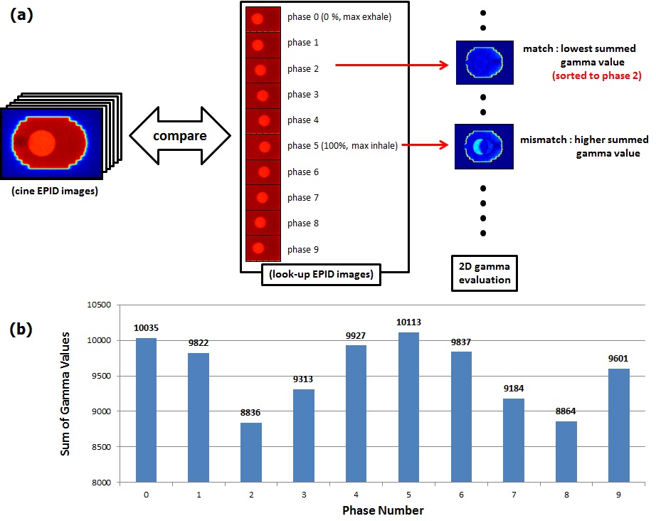
(1) A phantom, designed to simulate a tumor in lung (a polystyrene block with a 3 cm diameter embedded in cork), was placed on a sinusoidally moving platform with an amplitude of 1 cm and a period of 4 s. Ten-phase 4D computed tomography (CT) images of the phantom were acquired. A planning target volume (PTV) was created by adding a margin of 1 cm around the internal target volume of the tumor. (2) Three beams were designed, which included a static beam, a theoretical dynamic beam, and a planning-optimized dynamic beam (PODB). While the theoretical beam was made by manually programming a simplistic sliding leaf motion, the planning-optimized beam was obtained from treatment planning. From the three beams, three-dimensional (3D) doses on the phantom were calculated; 4D dose was calculated by means of the ten phase images (integrated over phases afterward); serving as "reference" images, phase-specific EPID dose images under the lung phantom were also calculated for each of the ten phases. (3) Cine EPID images were acquired while the beams were irradiated to the moving phantom. (4) Each cine image was phase-matched to a phase-specific CT image at which common irradiation occurred by intercomparing the cine image with the reference images. (5) Each cine image was used to reconstruct dose in the phase-matched CT image, and the reconstructed doses were summed over all phases. (6) The summation was compared with forwardly calculated 4D and 3D dose distributions. Accounting for realistic situations, intratreatment breathing irregularity was simulated by assuming an amplitude of 0.5 cm for the phantom during a portion of breathing trace in which the phase matching could not be performed. Intertreatment breathing irregularity between the time of treatment and the time of planning CT was considered by utilizing the same reduced amplitude when the phantom was irradiated. To examine the phase matching in a humanoid environment, the matching was also performed in a digital phantom (4D XCAT phantom).
RESULTS:
For the static, the theoretical, and the planning-optimized dynamic beams, the 4D reconstructed doses showed agreement with the forwardly calculated 4D doses within the gamma pass rates of 92.7%, 100%, and 98.1%, respectively, at the isocenter plane given by 3%/3 mm criteria. Excellent agreement in dose volume histogram of PTV and lung-PTV was also found between the two 4D doses, while substantial differences were found between the 3D and the 4D doses. The significant breathing irregularities modeled in this study were found not to be noticeably affecting the reconstructed dose. The phase matching was performed equally well in a digital phantom.
CONCLUSIONS:
The method of retrospective phase determination of a moving object under irradiation provided successful 4D dose reconstruction. This method will provide accurate quality assurance and facilitate adaptive therapy when distinguishable objects such as well-defined tumors, diaphragm, and organs with markers (pancreas and liver) are covered by treatment beam apertures.
Author information
Yoon J1, Jung JW1, Kim JO2, Yi BY3, Yeo I4.
1Department of Physics, East Carolina University, Greenville, North Carolina 27858.
2Department of Radiation Oncology, University of Pittsburgh Cancer Institute, Pittsburgh, Pennsylvania 15232.
3Department of Radiation Oncology, University of Maryland School of Medicine, Baltimore, Maryland 21201.
4Department of Radiation Medicine, Loma Linda University Medical Center, Loma Linda, California 92354.
- 연구소개
- 본 연구는 움직이는 기관에 흡수된 방사선량을 환자를 투과한 영상으로부터 재구성하는 방법을 제시하고, 이 방법을 임상에 적용하는 연구다. 각 방사선 조사시 움직이는 기관의 시간별 위치를 투과된 영상으로부터 찾아내어 해당 4D CT에 선량을 재구성한 것으로, 기관의 움직임이 불규칙할때도 정확하게 적용됨을 보인 연구이다. 미래의 방사선 조사는 움직임을 고려한 것으로, 이에 대한 선량투여 확인 및 적응형 치료(adaptive therapy)에, 현존하는 유일한 선량 역구성 방법으로서, 본 연구가 결정적으로 기여할 것으로 기대된다.
- 덧글달기


