글로벌 연구동향
의학물리학
![[Med Phys.] Three-dimensional quadratic modeling and quantitative evaluation of the diaphragm on a volumetric CT scan in patients with chronic obstructive pulmonary disease](/enewspaper/upimages/admin_20160811164031_R.png) [Med Phys.] Three-dimensional quadratic modeling and quantitative evaluation of the diaphragm on a volumetric CT scan in patients with chronic obstructive pulmonary disease
[Med Phys.] Three-dimensional quadratic modeling and quantitative evaluation of the diaphragm on a volumetric CT scan in patients with chronic obstructive pulmonary disease울산의대 / 장용준, 김남국*, 서준범*
- 출처
- Med Phys
- 등재일
- 2016 Jul
- 저널이슈번호
- 43(7):4273. doi: 10.1118/1.4953451.
- 내용
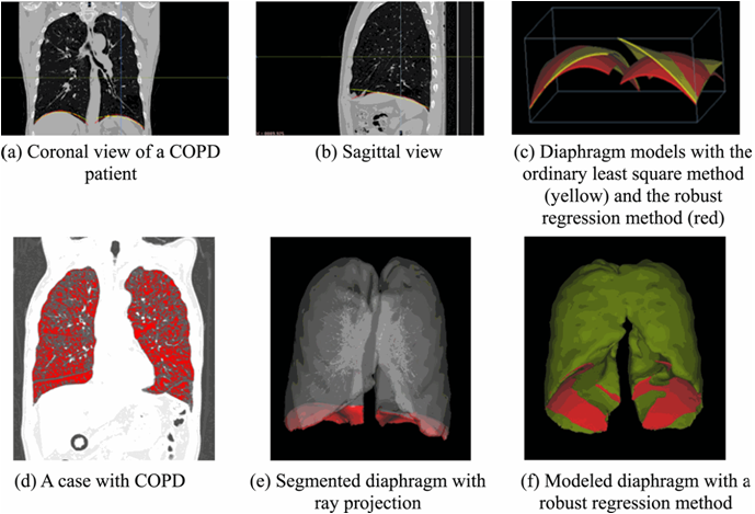
 Diaphragm segmentation and modeling with 3D quadratic surface fitting.
Abstract
PURPOSE:
In patients with chronic obstructive pulmonary disease (COPD), diaphragm function may deteriorate due to reduced muscle fiber length. Quantitative analysis of the morphology of the diaphragm is therefore important. In the authors current study, they propose a diaphragm segmentation method for COPD patients that uses volumetric chest computed tomography (CT) data, and they provide a quantitative analysis of the diaphragmatic dimensions.
METHODS:
Volumetric CT data were obtained from 30 COPD patients and 10 normal control patients using a 16-row multidetector CT scanner (Siemens Sensation 16) with 0.75-mm collimation. Diaphragm segmentation using 3D ray projections on the lower surface of the lungs was performed to identify the draft diaphragmatic lung surface, which was modeled using quadratic 3D surface fitting and robust regression in order to minimize the effects of segmentation error and parameterize diaphragm morphology. This result was visually evaluated by an expert thoracic radiologist. To take into consideration the shape features of the diaphragm, several quantification parameters-including the shape index on the apex (SIA) (which was computed using gradient set to 0), principal curvatures on the apex on the fitted diaphragm surface (CA), the height between the apex and the base plane (H), the diaphragm lengths along the x-, y-, and z-axes (XL, YL, ZL), quadratic-fitted diaphragm lengths on the z-axis (FZL), average curvature (C), and surface area (SA)-were measured using in-house software and compared with the pulmonary function test (PFT) results.
RESULTS:
The overall accuracy of the combined segmentation method was 97.22% ± 4.44% while the visual accuracy of the models for the segmented diaphragms was 95.28% ± 2.52% (mean ± SD). The quantitative parameters, including SIA, CA, H, XL, YL, ZL, FZL, C, and SA were 0.85 ± 0.05 (mm(-1)), 0.01 ± 0.00 (mm(-1)), 17.93 ± 10.78 (mm), 129.80 ± 11.66 (mm), 163.19 ± 13.45 (mm), 71.27 ± 17.52 (mm), 61.59 ± 16.98 (mm), 0.01 ± 0.00 (mm(-1)), and 34 380.75 ± 6680.06 (mm(2)), respectively. Several parameters were correlated with the PFT parameters.
CONCLUSIONS:
The authors propose an automatic method for quantitatively evaluating the morphological parameters of the diaphragm on volumetric chest CT in COPD patients. By measuring not only the conventional length and surface area but also the shape features of the diaphragm using quadratic 3D surface modeling, the proposed method is especially useful for quantifying diaphragm characteristics. Their method may be useful for assessing morphological diaphragmatic changes in COPD patients.
Author information
Chang Y1, Bae J2, Kim N3, Park JY4, Lee SM4, Seo JB4.
1School of Electrical Engineering, Korea Advanced Institute of Science and Technology, 291 Daehak-ro, Yuseong-gu, Daejeon 34138, South Korea.
2Interdisciplinary Program, Bioengineering Major, Graduate School, Seoul National University, Seoul 08826, South Korea.
3Department of Convergence Medicine, University of Ulsan College of Medicine, 88 Olympic-ro 43-gil, Songpa-gu, Seoul 05505, South Korea.
4Department of Radiology, University of Ulsan College of Medicine, 88 Olympic-ro 43-gil, Songpa-gu, Seoul 05505, South Korea.
- 연구소개
- 만성폐쇄성폐질환(COPD)의 영상 바이오마커인 횡경막의 정량적 측정과 폐기능검사와의 관련성을 분석한 논문입니다. Chest PA등에서 횡경막과 질환의 관계를 분석한 논문은 많으나, CT등에서 3차원으로 분석한 것은 많지 않습니다. CT에서 횡경막 분할이 쉽지 않기 때문에 자동화한 기법으로 횡경만을 분할하고, 이를 2차 함수로 모델링 하고, 정량화 한후에 COPD의 환자의 폐기능 검사와 관련성을 조사한 것입니다. 몇몇 정량지표는 FEV1, FEV1/FVC 등과 0.7 이상의 상관관계를 가지고 있는 것으로 밝혀졌습니다. 따라서 CT기반의 3차원 횡경막 정량지표가 COPD 등의 질환에서 환자의 severity를 반영하는 영상 바이오마커가 될 수 있다는 점을 의미한다고 할수 있겠습니다.
- 덧글달기


