


글로벌 연구동향
핵의학


![[Medicine (Baltimore).] Diagnostic value of 18F-FDG PET/CT in discriminating between benign and malignant lesions of the ribs](/enewspaper/upimages/1662341880admin.JPG) [Medicine (Baltimore).] Diagnostic value of 18F-FDG PET/CT in discriminating between benign and malignant lesions of the ribs
[Medicine (Baltimore).] Diagnostic value of 18F-FDG PET/CT in discriminating between benign and malignant lesions of the ribs울산의대 / 최선주, 김용일*
- 출처
- Medicine (Baltimore).
- 등재일
- 2022 Jul 8
- 저널이슈번호
- 101(27):e29867.
- 내용
Abstract
Purpose: Imaging biomarkers for rib mass are needed to optimize treatment plan. We investigated the diagnostic value of metabolic and volumetric parameters from 18F-fluorodeoxyglucose (FDG) positron-emission tomography/computed tomography (PET/CT) in discriminating between benign and malignant lesions of the ribs.Patients and methods: Fifty-seven patients with pathologically proven diagnosis of rib lesions were retrospectively enrolled. The size of rib lesions, the maximum, mean, and peak standardized uptake value (SUVmax, SUVmean, SUVpeak), tumor-to-background ratio (TBR), metabolic tumor volume (MTV), and total lesions glycolysis (TLG) were measured. The FDG uptake patterns (segmental and discrete) and CT findings (soft tissue involvement and fracture) were also reviewed.
Results: Among the multiple parameters extracted from PET/CT, the MTV of malignant lesions was significantly higher than that of benign lesions (median; 4.7 vs 0.2, respectively, P = .041). In receiver operating characteristics curve analysis, MTV had the largest area under curve of 0.672 for differentiating malignant from benign lesions. For identifying malignant lesions, an MTV threshold of 0.5 had a sensitivity of 85.0%, specificity of 47.1%, positive predictive value of 79.1%, negative predictive value of 57.1%, and accuracy of 73.7%. The presence of adjacent soft tissue involvement around rib lesions showed a significant association with malignancy (odds ratio = 6.750; 95% CI, 1.837-24.802, P = .003).
Conclusions: The MTV is a useful PET/CT parameter for assisting in the differential diagnosis of suspected malignant lesions of the ribs. CT finding of adjacent soft tissue involvement around rib was significantly associated with malignant lesions of the ribs.
Affiliations
Sunju Choi 1 2 , Yong-Il Kim 1 , Geun Dong Lee 3 , Sehoon Choi 3 , Hyeong Ryul Kim 3 , Yong-Hee Kim 3 , Dong Kwan Kim 3 , Seung-Il Park 3 , Jin-Sook Ryu 1
1 Department of Nuclear Medicine, Asan Medical Center, University of Ulsan college of Medicine, Seoul, Republic of Korea.
2 Department of Nuclear Medicine, Kyung Hee University Hospital, Kyung Hee University School of Medicine, Seoul, Republic of Korea.
3 Department of Thoracic and Cardiovascular Surgery, Asan Medical Center, University of Ulsan College of Medicine, Seoul, Republic of Korea.
- 키워드
- TLICS; bone scan; burst fracture; scintigraphy; unstable.
- 연구소개
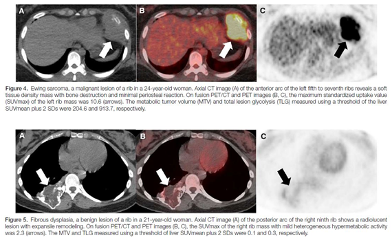
- Rib lesion의 치료 방침을 결정하기 위한 imaging biomarker로서 F-18-FDG PET/CT의 진단적 성능을 알아보기 위한 연구입니다. Primary rib malignancy (그림 1), rib metastasis, 그리고 benign rib lesion (그림 2) 들의 FDG uptake와 관련된 정성적, 정량적 특성들과 CT finding들을 비교하였고, 치료 방침을 결정하는데 F-18-FDG PET/CT의 metabolic tumor volume이 도움이 될 수 있음을 알 수 있었습니다.
- 덧글달기


![]()
- COPYRIGHT(C) 2015 한국원자력의학원 전략기획팀 All rights Reserved.
- 문의 : rmwebzine@kirams.re.kr 발행처 : 한국원자력의학원 전략기획팀
- 우) 01812 서울시 노원구 노원로 75 한국원자력의학원 전략기획팀