


글로벌 연구동향
방사선종양학


![[PLoS One .] Assessment of lymph node area coverage with total marrow irradiation and implementation of total marrow and lymphoid irradiation using automated deep learning-based segmentation](/enewspaper/upimages/1711954961admin.JPG) [PLoS One .] Assessment of lymph node area coverage with total marrow irradiation and implementation of total marrow and lymphoid irradiation using automated deep learning-based segmentation
[PLoS One .] Assessment of lymph node area coverage with total marrow irradiation and implementation of total marrow and lymphoid irradiation using automated deep learning-based segmentation서울의대 / 최현석, 장범섭*, 장지현*
- 출처
- PLoS One .
- 등재일
- 2024 Mar 8
- 저널이슈번호
- 19(3):e0299448.
- 내용
Abstract
Background: Total marrow irradiation (TMI) and total marrow and lymphoid irradiation (TMLI) have the advantages. However, delineating target lesions according to TMI and TMLI plans is labor-intensive and time-consuming. In addition, although the delineation of target lesions between TMI and TMLI differs, the clinical distinction is not clear, and the lymph node (LN) area coverage during TMI remains uncertain. Accordingly, this study calculates the LN area coverage according to the TMI plan. Further, a deep learning-based model for delineating LN areas is trained and evaluated.Methods: Whole-body regional LN areas were manually contoured in patients treated according to a TMI plan. The dose coverage of the delineated LN areas in the TMI plan was estimated. To train the deep learning model for automatic segmentation, additional whole-body computed tomography data were obtained from other patients. The patients and data were divided into training/validation and test groups and models were developed using the "nnU-NET" framework. The trained models were evaluated using Dice similarity coefficient (DSC), precision, recall, and Hausdorff distance 95 (HD95). The time required to contour and trim predicted results manually using the deep learning model was measured and compared.
Results: The dose coverage for LN areas by TMI plan had V100% (the percentage of volume receiving 100% of the prescribed dose), V95%, and V90% median values of 46.0%, 62.1%, and 73.5%, respectively. The lowest V100% values were identified in the inguinal (14.7%), external iliac (21.8%), and para-aortic (42.8%) LNs. The median values of DSC, precision, recall, and HD95 of the trained model were 0.79, 0.83, 0.76, and 2.63, respectively. The time for manual contouring and simply modified predicted contouring were statistically significantly different.
Conclusions: The dose coverage in the inguinal, external iliac, and para-aortic LN areas was suboptimal when treatment is administered according to the TMI plan. This research demonstrates that the automatic delineation of LN areas using deep learning can facilitate the implementation of TMLI.
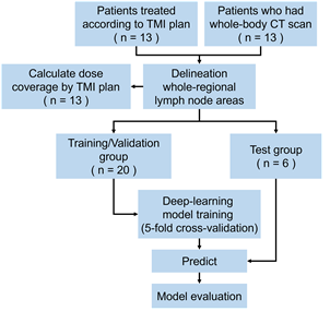
연구 schema
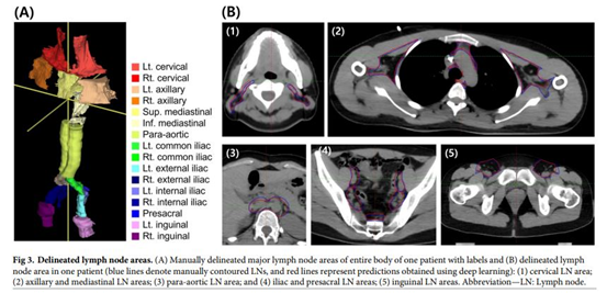
(A) 수동으로 구획된 전신 림프절 영역 (B) 푸른선 : 수동으로 구획된 림프절영역, 붉은선: 딥러닝을 통해 구획된 림프절영역
Affiliations
Hyeon Seok Choi 1 2, Hyun-Cheol Kang 1 2, Eui Kyu Chie 1 2, Kyung Hwan Shin 1 2, Ji Hyun Chang 1 2, Bum-Sup Jang 1 2
1Department of Radiation Oncology, Seoul National University Hospital, Seoul, South Korea.
2Department of Radiation Oncology, Seoul National University College of Medicine, Seoul, South Korea.
- 연구소개
- 전체 골수 조사(TMI) 계획에 따라 림프절 영역의 복사선 피폭 범위를 분석하고, 림프절 영역을 자동으로 구분하기 위한 딥러닝 모델을 개발 및 평가한 논문입니다. 현재 임상에서 쓰이고 있는 치료를 평가하고, 보완하기 위한 방법으로 딥러닝을 사용하였습니다. 이를 통해 방사선의학 분야에 있어 딥러닝을 어떻게 사용하면 좋을지, 그리고 딥러닝 모델을 어떤 방식으로 평가하는지 잘 모를 수 있는 연구자들에게 도움이 될 수 있는 정보라고 생각합니다.
- 덧글달기


![]()
- COPYRIGHT(C) 2015 한국원자력의학원 전략기획팀 All rights Reserved.
- 문의 : rmwebzine@kirams.re.kr 발행처 : 한국원자력의학원 전략기획팀
- 우) 01812 서울시 노원구 노원로 75 한국원자력의학원 전략기획팀