


글로벌 연구동향
방사선종양학


![[Cancers (Basel).] Impact of Denoising on Deep-Learning-Based Automatic Segmentation Framework for Breast Cancer Radiotherapy Planning](/enewspaper/upimages/1662339282admin.JPG) [Cancers (Basel).] Impact of Denoising on Deep-Learning-Based Automatic Segmentation Framework for Breast Cancer Radiotherapy Planning
[Cancers (Basel).] Impact of Denoising on Deep-Learning-Based Automatic Segmentation Framework for Breast Cancer Radiotherapy Planning연세의대 / 임정호, 이익재, 이호*
- 출처
- Cancers (Basel).
- 등재일
- 2022 Jul 22
- 저널이슈번호
- 14(15):3581. doi: 10.3390/cancers14153581.
- 내용
Abstract
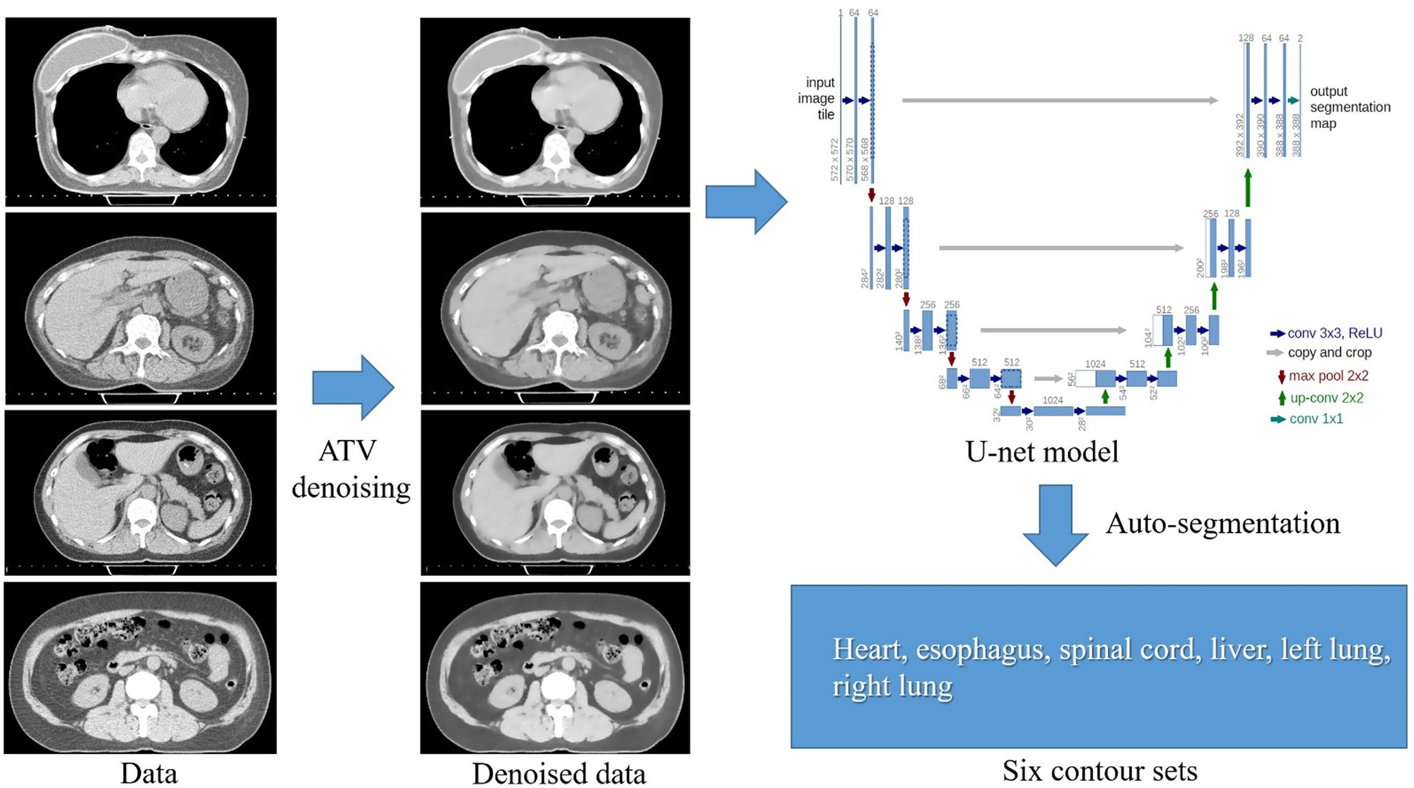
Objective: This study aimed to investigate the segmentation accuracy of organs at risk (OARs) when denoised computed tomography (CT) images are used as input data for a deep-learning-based auto-segmentation framework.Methods: We used non-contrast enhanced planning CT scans from 40 patients with breast cancer. The heart, lungs, esophagus, spinal cord, and liver were manually delineated by two experienced radiation oncologists in a double-blind manner. The denoised CT images were used as input data for the AccuContourTM segmentation software to increase the signal difference between structures of interest and unwanted noise in non-contrast CT. The accuracy of the segmentation was assessed using the Dice similarity coefficient (DSC), and the results were compared with those of conventional deep-learning-based auto-segmentation without denoising.
Results: The average DSC outcomes were higher than 0.80 for all OARs except for the esophagus. AccuContourTM-based and denoising-based auto-segmentation demonstrated comparable performance for the lungs and spinal cord but showed limited performance for the esophagus. Denoising-based auto-segmentation for the liver was minimal but had statistically significantly better DSC than AccuContourTM-based auto-segmentation (p < 0.05).
Conclusions: Denoising-based auto-segmentation demonstrated satisfactory performance in automatic liver segmentation from non-contrast enhanced CT scans. Further external validation studies with larger cohorts are needed to verify the usefulness of denoising-based auto-segmentation.
저잡음 필터링을 이용한 딥러닝기반 자동분할 워크플로우
Affiliations
Jung Ho Im 1 , Ik Jae Lee 2 , Yeonho Choi 3 , Jiwon Sung 2 , Jin Sook Ha 3 , Ho Lee 2
1 CHA Bundang Medical Center, Department of Radiation Oncology, CHA University School of Medicine, Seongnam 13496, Korea.
2 Department of Radiation Oncology, Yonsei University College of Medicine, Seoul 03722, Korea.
3 Department of Radiation Oncology, Gangnam Severance Hospital, Seoul 06273, Korea.
- 키워드
- contouring; deep-learning-based auto-segmentation; denoiser; organs at risk; radiation therapy.
- 연구소개
- 딥러닝기반 학습모델이 탑재된 자동분할소프트웨어에 입력데이터로 저잡음 필터링을 통해 획득된 비조영 CT영상이 주어졌을 때 organs at risk 분할 정확도를 조사한 논문입니다. 유방암 환자 40명 대상으로 저잡음 필터링 적용한 경우와 그렇지 않은 경우를 비교한 결과, Lung과 spinal cord 분할에 대해서는 유사한 성능을 보여주었지만, 간 분할에서는 저잡음 필터링이 통계적으로 유의하게 우수한 성능을 보였습니다. 저잡음 필터링을 딥러닝 학습모델과 결합할 때 간 분할 정확도를 향상시킬 수 있는 가능성을 시사하고 있습니다.
- 덧글달기
- 이전글 [Front Oncol.] Trends in radiotherapy administration in the management of hepatocellular carcinoma: Analysis of a Korean tertiary hospital registry of hepatocellular carcinoma patients diagnosed between 2005 and 2017
- 다음글 [Cancers (Basel).] Proton Beam Therapy versus Photon Radiotherapy for Stage I Non-Small Cell Lung Cancer


![]()
- COPYRIGHT(C) 2015 한국원자력의학원 전략기획팀 All rights Reserved.
- 문의 : rmwebzine@kirams.re.kr 발행처 : 한국원자력의학원 전략기획팀
- 우) 01812 서울시 노원구 노원로 75 한국원자력의학원 전략기획팀