(구)글로벌 핫이슈
방사선종양학
- 2017년 04월호
[J Clin Oncol] Cetuximab Plus Chemoradiotherapy in Immunocompetent Patients With Anal Carcinoma: A Phase II Eastern Cooperative Oncology Group-American College of Radiology Imaging Network Cancer Research Group Trial (E3205).Montefiore Medical Center / Joseph A. Sparano*
- 출처
- J Clin Oncol
- 등재일
- 2017 Mar
- 저널이슈번호
- 35(7):718-726. doi: 10.1200/JCO.2016.69.1667. Epub 2017 Jan 9.
- 내용
Abstract
Purpose Squamous cell carcinoma of the anal canal (SCCAC) is characterized by high locoregional failure (LRF) rates after sphincter-preserving definitive chemoradiation (CRT) and is typically associated with anogenital human papilloma virus infection. Because cetuximab enhances the effect of radiation therapy in human papilloma virus-associated oropharyngeal squamous cell carcinoma, we hypothesized that adding cetuximab to CRT would reduce LRF in SCCAC. Methods Sixty-one patients with stage I to III SCCAC received CRT including cisplatin, fluorouracil, and radiation therapy to the primary tumor and regional lymph nodes (45 to 54 Gy) plus eight once-weekly doses of concurrent cetuximab. The study was designed to detect at least a 50% reduction in 3-year LRF rate (one-sided α, 0.10; power 90%), assuming a 35% LRF rate from historical data. Results Poor risk features included stage III disease in 64% and male sex in 20%. The 3-year LRF rate was 23% (95% CI, 13% to 36%; one-sided P = .03) by binomial proportional estimate using the prespecified end point and 21% (95% CI, 7% to 26%) by Kaplan-Meier estimate in a post hoc analysis using methods consistent with historical data. Three-year rates were 68% (95% CI, 55% to 79%) for progression-free survival and 83% (95% CI, 71% to 91%) for overall survival. Grade 4 toxicity occurred in 32%, and 5% had treatment-associated deaths. Conclusion Although the addition of cetuximab to chemoradiation for SCCAC was associated with lower LRF rates than historical data with CRT alone, toxicity was substantial, and LRF still occurs in approximately 20%, indicating the continued need for more effective and less toxic therapies.

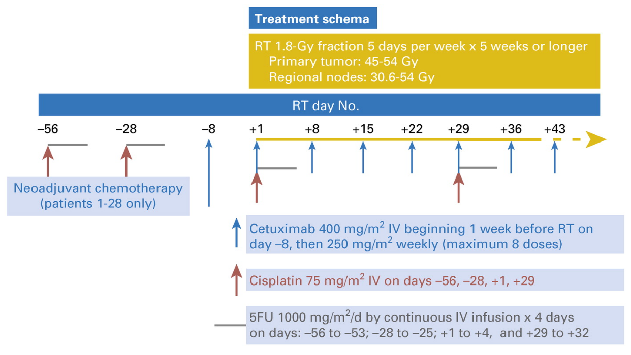
Fig 1.
Treatment schema. FU, fluorouracil; IV, intravenous; RT, radiation therapy.
Author information
Garg MK1, Zhao F1, Sparano JA1, Palefsky J1, Whittington R1, Mitchell EP1, Mulcahy MF1, Armstrong KI1, Nabbout NH1, Kalnicki S1, El-Rayes BF1, Onitilo AA1, Moriarty DJ1, Fitzgerald TJ1, Benson AB 3rd1.
1 Madhur K. Garg, Joseph A. Sparano, and Shalom Kalnicki, Montefiore Medical Center, Albert Einstein College of Medicine, Montefiore-Einstein Center for Cancer Care, Bronx, NY; Fengmin Zhao, Dana Farber Cancer Institute, Boston, MA; Joel Palefsky, University of California, San Francisco, CA; Richard Whittington, Philadelphia VA Medical Center; Edith P. Mitchell, Thomas Jefferson University, Philadelphia, PA; Mary F. Mulcahy and Al B. Benson III, Northwestern University, Chicago, IL; Karin I. Armstrong, United Hospital, Woodbury, MN; Nassim H. Nabbout, Cancer Center of Kansas, Wichita, KS; Bassel F. El-Rayes, Emory University, Atlanta, GA; Adedayo A. Onitilo, Marshfield Clinic, Marshfield, WI; Daniel J. Moriarty, Overlook Medical Center, Summit, NJ; and Thomas J. Fitzgerald, Imaging and Radiation Oncology Core, Quality Assurance Review Center, Providence, RI.
- 덧글달기
- 이전글 [J Clin Oncol. ] Cetuximab Plus Chemoradiotherapy for HIV-Associated Anal Carcinoma: A Phase II AIDS Malignancy Consortium Trial.
- 다음글 [Lancet Oncol.] Best time to assess complete clinical response after chemoradiotherapy in squamous cell carcinoma of the anus (ACT II): a post-hoc analysis of randomised controlled phase 3 trial.


