


글로벌 연구동향
분자영상 및 방사화학


![[Front Vet Sci.] Case Report: 18F-Fluoro-L-Phenylalanine Positron Emission Tomography Findings and Immunoreactivity for L-Type Amino Acid Transporter 1 in a Dog With Meningioma](/enewspaper/upimages/1662342615admin.JPG) [Front Vet Sci.] Case Report: 18F-Fluoro-L-Phenylalanine Positron Emission Tomography Findings and Immunoreactivity for L-Type Amino Acid Transporter 1 in a Dog With Meningioma
[Front Vet Sci.] Case Report: 18F-Fluoro-L-Phenylalanine Positron Emission Tomography Findings and Immunoreactivity for L-Type Amino Acid Transporter 1 in a Dog With Meningioma충북대 / 이도희, 강병택*
- 출처
- Front Vet Sci.
- 등재일
- 2022 Jul 15
- 저널이슈번호
- 9:899229. doi: 10.3389/fvets.2022.899229.
- 내용
Abstract
A 12-year-old intact female Miniature Pinscher dog weighing 5.4 kg presented with a history of seizures. On neurological examination, postural reactions were decreased in the left-sided limbs, and menace responses were bilaterally absent. Magnetic resonance imaging (MRI) of the brain was performed, and a solitary amorphous mass (2.7 × 1.9 × 2.2 cm) was observed on the right side of the frontal lobe. Based on the signalment, clinical signs, and MRI findings, a brain tumor was tentatively diagnosed, and meningioma was suspected. The dog was treated with hydroxyurea, prednisolone, and other antiepileptic drugs. One week after the treatment began, postural reactions returned to normal, and the menace response improved. At 119 days after treatment, 18F-fluoro-L-phenylalanine (18F-FDOPA) positron emission tomography (PET) was performed. Marked 18F-FDOPA uptake was observed in the lesion. The mean and maximal standardized uptake values of the lesion were 2.61 and 3.72, respectively, and the tumor-to-normal tissue ratio was 1.95. At 355 days after the initial treatment, a second MRI scan was performed and the tumor size had increased to 3.5 × 2.8 × 2.9 cm. The dog died 443 days after the initial treatment and was definitively diagnosed with grade 1 meningioma by histopathological examination. Immunohistochemical staining for Ki67 and L-type amino acid transporter 1 was positive and negative for p53, respectively. The labeling index of Ki67 was 2.4%. This is the first case to demonstrate 18F-FDOPA PET findings in a clinical case of a dog histologically diagnosed with a meningioma.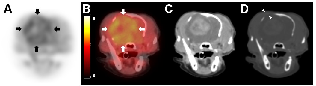
Affiliations
Dohee Lee 1 , Taesik Yun 1 , Sanggu Kim 2 , Yoonhoi Koo 1 , Yeon Chae 1 , Soochong Kim 2 , Dongwoo Chang 3 , Mhan-Pyo Yang 1 , Hakhyun Kim 1 , Byeong-Teck Kang 1
1 Laboratory of Veterinary Internal Medicine, College of Veterinary Medicine, Chungbuk National University, Cheongju, South Korea.
2 Laboratory of Veterinary Pathology and Platelet Signaling, College of Veterinary Medicine, Chungbuk National University, Cheongju, South Korea.
3 Department of Veterinary Imaging, College of Veterinary Medicine, Chungbuk National University, Cheongju, South Korea.
- 키워드
- 18F-FDOPA; L-type amino acid transporter 1; brain tumor; canine; meningioma; positron emission tomography.
- 연구소개
- 개에서 가장 흔한 뇌종양 중 하나인 뇌수막종 (meningioma)에 이환된 12살령 미니핀 개에서 18F-FDOPA PET finding에 대한 케이스 보고입니다. 18F-FDOPA는 amino acid analog로, 사람에서 뇌종양의 진단에 대한 민감도가 glucose analog인 18F-FDG보다 월등히 높다 알려져 있습니다. 그러나 수의학에서 뇌종양 환자에 대한 18F-FDOPA 적용 보고는 매우 제한적이며, 개의 신경아교종 (glioma)에서 적용에 대한 보고가 유일합니다. 따라서 이 논문은 개 뇌수막종에서 18F-FDOPA PET 적용에 대한 최초 보고입니다. 또한 본 케이스는 개 뇌수막종 조직에서 L-type amino acid transporter 1 (LAT1)에 대한 면역조직화학염색을 시행함으로써, LAT1이 진단적 PET imaging을 위한 molecular target이 될 수 있음을 시사합니다.
- 덧글달기


![]()
- COPYRIGHT(C) 2015 한국원자력의학원 전략기획팀 All rights Reserved.
- 문의 : rmwebzine@kirams.re.kr 발행처 : 한국원자력의학원 전략기획팀
- 우) 01812 서울시 노원구 노원로 75 한국원자력의학원 전략기획팀