


글로벌 연구동향
분자영상 및 방사화학


![[Eur J Nucl Med Mol Imaging.] In vivo detection of hydrogen sulfide in the brain of live mouse: application in neuroinflammation models](/enewspaper/upimages/1659510420admin.PNG) [Eur J Nucl Med Mol Imaging.] In vivo detection of hydrogen sulfide in the brain of live mouse: application in neuroinflammation models
[Eur J Nucl Med Mol Imaging.] In vivo detection of hydrogen sulfide in the brain of live mouse: application in neuroinflammation modelsKIRAMS, 경북대 / 남보라, 박현, 김정영, 이교철, 석경호, 유정수*
- 출처
- Eur J Nucl Med Mol Imaging.
- 등재일
- 2022 Jun 10. doi: 10.1007/s00259-022-05854-1.
- 저널이슈번호
- 내용
Abstract
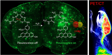
Purpose: Hydrogen sulfide (H2S) plays important roles in brain pathophysiology. However, nuclear imaging probes for the in vivo detection of brain H2S in living animals have not been developed. Here, we report the first nuclear imaging probe that enables in vivo imaging of endogenous H2S in the brain of live mice.Methods: Utilizing a bis(thiosemicarbazone) backbone, a fluorescent ATSM-FITC conjugate was synthesized. Its copper complex, Cu(ATSM-FITC) was thoroughly tested as a biosensor for H2S. The same ATSM-FITC ligand was quantitatively labeled with [64Cu]CuCl2 to obtain a radioactive [64Cu][Cu(ATSM-FITC)] imaging probe. Biodistribution and positron emission tomography (PET) imaging studies were performed in healthy mice and neuroinflammation models.
Results: The Cu(ATSM-FITC) complex reacts instantly with H2S to release CuS and becomes fluorescent. It showed excellent reactivity, sensitivity, and selectivity to H2S. Endogenous H2S levels in living cells were successfully detected by fluorescence microscopy. Exceptionally high brain uptake of [64Cu][Cu(ATSM-FITC)] (> 9% ID/g) was observed in biodistribution and PET imaging studies. Subtle changes in brain H2S concentrations in live mice were accurately detected by quantitative PET imaging. Due to its dual modality feature, increased H2S levels in neuroinflammation models were characterized at the subcellular level by fluorescence imaging and at the whole-body scale by PET imaging.
Conclusion: Our biosensor can be readily utilized to study brain H2S function in live animal models and shows great potential as a novel imaging agent for diagnosing brain diseases.
Affiliations
Bora Nam # 1 , Woonghee Lee # 1 , Swarbhanu Sarkar # 1 , Jae-Hong Kim 2 , Abhinav Bhise 1 , Hyun Park 3 , Jung Young Kim 3 , Phuong Tu Huynh 1 , Subramani Rajkumar 1 , Kiwoong Lee 1 , Yeong Su Ha 1 , Seong Hwan Cho 1 , Jeong Eun Lim 1 , Kyung Won Kim 1 , Kyo Chul Lee 3 , Kyoungho Suk 2 , Jeongsoo Yoo 4
1 Department of Molecular Medicine, Brain Korea 21 four KNU Convergence Educational Program of Biomedical Sciences for Creative Future Talents, School of Medicine, Kyungpook National University, Daegu, 41944, South Korea.
2 Department of Pharmacology, Brain Korea 21 four KNU Convergence Educational Program of Biomedical Sciences for Creative Future Talents, School of Medicine, Brain Science & Engineering Institute, Kyungpook National University, Daegu, 41944, South Korea.
3 Division of Applied RI, Korea Institute of Radiological and Medical Sciences, Seoul, 01812, South Korea.
4 Department of Molecular Medicine, Brain Korea 21 four KNU Convergence Educational Program of Biomedical Sciences for Creative Future Talents, School of Medicine, Kyungpook National University, Daegu, 41944, South Korea. yooj@knu.ac.kr.
# Contributed equally.
- 키워드
- Gasotransmitter; Hydrogen sulfide; Imaging agent; Neuroinflammation.
- 연구소개
- 황화수소(H2S)는 뇌 안에서 다양한 생리/병리작용에 관여하고 있지만 이를 효과적으로 모니터링할 수 있는 임상적 방법은 전무한 상황입니다. 본 연구에서는 뇌의 혈액뇌관문(Blood-brain barrier)를 매우 잘 통과하면서 황화수소하고만 높은 민감도로 선택적 반응을 하는 방사성 프로브를 새롭게 개발하였습니다. 많은 뇌 질환에서 뇌염증이 수반되고 이 경우 황화수소의 농도도 증가된다는 사실에 착안하여, 뇌염증 동물모델에서 뇌염증 부위를 PET 영상을 통해 성공적으로 진단할 수 있었습니다. 저희가 개발한 프로브가 향후 뇌염증을 동반한 다양한 뇌질환의 조기 진단에 활용될 수 있기를 기대해 봅니다.
- 덧글달기


![]()
- COPYRIGHT(C) 2015 한국원자력의학원 전략기획팀 All rights Reserved.
- 문의 : rmwebzine@kirams.re.kr 발행처 : 한국원자력의학원 전략기획팀
- 우) 01812 서울시 노원구 노원로 75 한국원자력의학원 전략기획팀