글로벌 연구동향
분자영상 및 방사화학
![[Biomaterials.] Targeted multimodal nano-reporters for pre-procedural MRI and intra-operative image-guidance.](/enewspaper/upimages/admin_20170111140550_R.png) 2017년 01월호
2017년 01월호
[Biomaterials.] Targeted multimodal nano-reporters for pre-procedural MRI and intra-operative image-guidance.Northwestern University / Elena A. Rozhkova*,김동현*
- 출처
- Biomaterials.
- 등재일
- 2016 Dec
- 저널이슈번호
- 109:69-77. doi: 10.1016/j.biomaterials.2016.09.013. Epub 2016 Sep 19.
- 내용

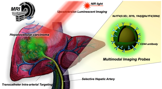
그림1) Multimodal MR/Upconversion luminescent imaging of HCC tumor using transcatheter hepatic intra-arterial (IA) targeted anti-CD44-Nd-CSUCNPs. Multi-modal imaging reporters, Nd-CSUCNPs conjugated with an anti-CD44 monoclonal antibody, were delivered intra-arterially for the sensitive and rapid detection of HCC in orthotopic HCC rat models. IA delivery of multi-modality Nd-CSUCNPs conjugated with specific tumor targeting molecules facilitates rapid and selective targeting of Nd-CSUCNPs to HCC, providing strong image-contrast between the tumors and normal hepatic tissue. Next, the feasibility of multimodal diagnostic MR and intraoperative UCL image-guided surgery for HCC resection was demonstrated.
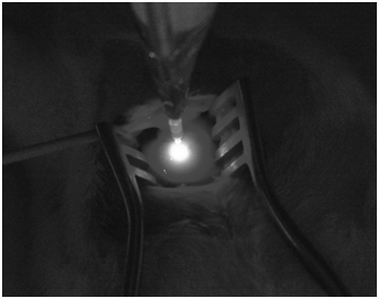
그림2) An luminescent image of liver tumor in a HCC rat with 808 NIR laser and targeted anti-CD44 UCNPs.Abstract
Multimodal-imaging probes offer a novel approach, which can provide detail diagnostic information for the planning of image-guided therapies in clinical practice. Here we report targeted multimodal Nd3+-doped upconversion nanoparticle (UCNP) imaging reporters, integrating both magnetic resonance imaging (MRI) and real-time upconversion luminescence imaging (UCL) capabilities within a single platform. Nd3+-doped UCNPs were synthesized as a core-shell structure showing a bright visible emission upon excitation at the near infrared (minimizing biological overheating and increasing tissue penetration depth) as well as providing strong MRI T2 contrast (high r2/r1 ratio). Transcatheter intra-arterial infusion of Nd3+-doped UCNPs conjugated with anti-CD44-monoclonal antibody allowed for high performance in vivo multimodal UCL and MR imaging of hepatocellular carcinoma (HCC) in an orthotopic rat model. The resulted in vivo multimodal imaging of Nd3+ doped core-shell UCNPs combined with transcatheter intra-arterial targeting approaches successfully discriminated liver tumors from normal hepatic tissues in rats for surgical resection applications. The demonstrated multimodal UCL and MRI imaging capabilities of our multimodal UCNPs reporters suggest strong potential for in vivo visualization of tumors and precise surgical guidance to fill the gap between pre-procedural imaging and intraoperative reality.
Author information
Lee J1, Gordon AC2, Kim H3, Park W4, Cho S4, Lee B5, Larson AC6, Rozhkova EA7, Kim DH8.
1Center for Nanoscale Materials, Argonne National Laboratory, Argonne, IL 60439, USA.
2Department of Radiology, Northwestern University Feinberg School of Medicine, Chicago, IL 60611, USA; Department of Biomedical Engineering, Northwestern University, Evanston, IL 60208, USA.
3Department of Chemistry, Northwestern University, Evanston, IL 60208, USA.
4Department of Radiology, Northwestern University Feinberg School of Medicine, Chicago, IL 60611, USA.
5Advanced Photon Source, Argonne National Laboratory, Argonne, IL 60439, USA.
6Department of Radiology, Northwestern University Feinberg School of Medicine, Chicago, IL 60611, USA; Robert H. Lurie Comprehensive Cancer Center, Chicago, IL 60611, USA; Department of Biomedical Engineering, Northwestern University, Evanston, IL 60208, USA; Department of Electrical Engineering and Computer Science, Evanston, IL 60208, USA; International Institute of Nanotechnology (IIN), Northwestern University, Evanston, IL 60208, USA.
7Center for Nanoscale Materials, Argonne National Laboratory, Argonne, IL 60439, USA. Electronic address: rozhkova@anl.gov.
8Department of Radiology, Northwestern University Feinberg School of Medicine, Chicago, IL 60611, USA; Robert H. Lurie Comprehensive Cancer Center, Chicago, IL 60611, USA. Electronic address: dhkim@northwestern.edu.
- 키워드
- Cancer; Interventional radiology; Medical imaging; Multimodal probe; Upconversion nanoparticles
- 연구소개
- 의료 영상기술은 현재 사용되고 있는 다양한 질병 치료법과 함께 진단과 예후 판단을 위해 계속적인 발전 중에 있는 분야이다. 항암 치료 분야에서 보자면, 임상에서 환자의 암의 진행 단계에 따른 다양한 치료방법이 사용되고 있다. 특히 병변 부위를 외과적으로 절개하여 암을 제거하는 외과적 절제술을 주로 사용하고 있는 실정이다. 하지만, 암 병변 절제시 시술자의 경험에 의해 암조직과 주변조직의 경계가 판단되어 제거가 수행되므로 남아있는 암 조직이 재발될 수 있어, 그에 대한 예후가 좋지 않다. 따라서 수술 중 병변 부위를 정확히 영상화할 수 있는 기술이 시급한 실정이다. 본 연구에서는 이러한 수술 전/수술 중/수술 후의 모든 단계에서 병변 부위를 정확히 영상화할 수 있는 나노 입자를 개발하여 그 가능성을 동물 실험을 통해 입증하였다. 자기공명 영상(MRI) 조영제인 산화철 나노 입자, 수술 중 암 조직 영상화에 사용되어온 형광 프로브(예; Indocyanin green)들이 현재 임상에서 외과적 암 수술을 위한 각 단계에서 사용할 수 있는 것들이다. 하지만, 암 표적률이 높지 않고, 각 단계마다 다른 종류의 조영제를 써야 하는 단점들로 인해 그 사용이 제한되어 있다. 본 연구에서는 자기공명 (MR)과 발광(Luminescent) 영상 장치를 통해 조영이 가능한 업컨버전 나노입자를 개발하여 수술 시 정확히 병변 부위를 영상화하여 시술자가 병변 부위를 완벽히 제거할 수 있도록 하였다. 개발된 Nd계열의 업컨버전 나노입자는 MRI T1조영은 물론 인체조직 투과율이 높은 근적외선 광을 이용하여 밝은 빛을 내는 다기능성이 입증되었다. 그에 더하여 암을 표적화할 수 있는 특정 항체를 표면에 접합하여 암 표적률을 향상시켰다. 따라서 본 연구에서 개발된 나노입자는 향후 다양한 암 치료에 이용될 수 있을 것이라 기대할 수 있다.
- 덧글달기


