글로벌 연구동향
의학물리학
![[Radiother Oncol.] Two-dimensional in vivo rectal dosimetry using an endorectal balloon with unfoldable radiochromic film during prostate cancer radiotherapy.](/enewspaper/upimages/admin_20160609142424_R.bmp) 2016년 06월호
2016년 06월호
[Radiother Oncol.] Two-dimensional in vivo rectal dosimetry using an endorectal balloon with unfoldable radiochromic film during prostate cancer radiotherapy.국립암센터/ 정은희, 민순기, 임영경*
- 출처
- Radiother Oncol.
- 등재일
- 2016 May 21
- 저널이슈번호
- pii: S0167-8140(16)31091-X. doi: 10.1016/j.radonc.2016.05.003.
- 내용

Abstract
BACKGROUND AND PURPOSE:
The present study aims to investigate the feasibility of two-dimensional (2D) in vivo rectal dosimetry using an endorectal balloon for the radiotherapy of prostate cancer.
MATERIALS AND METHODS:
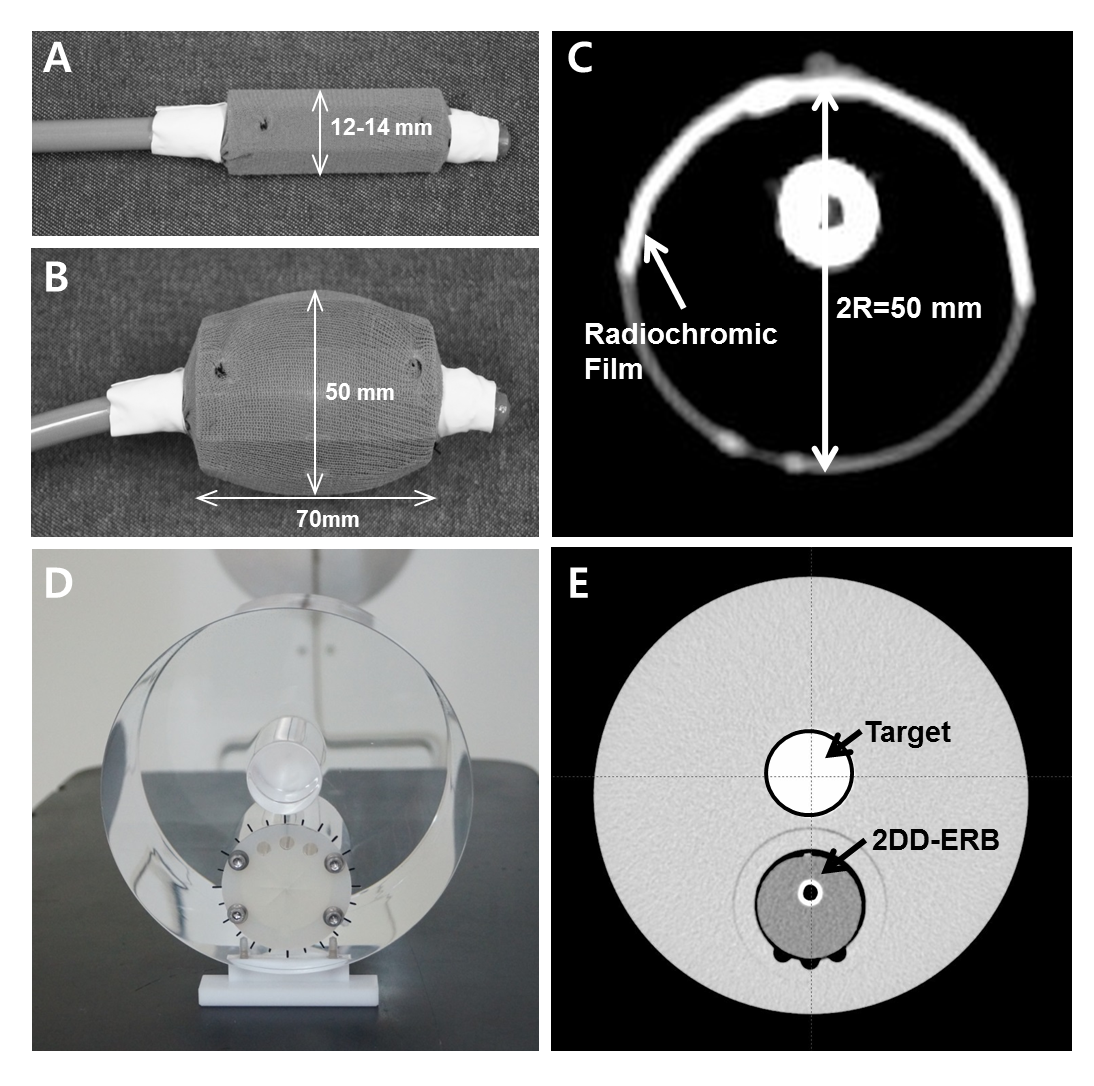
The endorectal balloon was equipped with an unfoldable radiochromic film. The film was unrolled as the balloon was inflated, and rolled as it was deflated. Its mechanical and imaging properties were tested, and the dosimetric effectiveness was evaluated in clinical photon and proton beams.
RESULTS:
The size of the endorectal balloon including the film was linearly proportional to the volume of water filled in the balloon, and its position could be identified by X-ray radiography. The loss of dose information due to film cutting was within ±1mm from the cutting line. Applying linear interpolation on cut film, the gamma passing rate was more than 95% for 2%/2mm criteria. The measured dose profiles agreed with the plan within 3% and 4% for the photon and proton beams, respectively. A dose-volume histogram of the anterior rectal wall could be obtained from the measured dose distribution in the balloon, which also agreed well with the plan.
CONCLUSIONS:
2D in vivo rectal dosimetry is feasible using the endorectal balloon with a radiochromic film in the radiotherapy of prostate cancer.
Author information
Jeang EH1, Min S1, Cho KH1, Hwang UJ2, Choi SH3, Kwak J4, Jeong JH1, Kim H1, Lee SB1, Shin D1, Park J1, Kim JY1, Kim DY1, Lim YK5.
1Proton Therapy Center, National Cancer Center, Goyang, Republic of Korea.
2Department of Radiation Oncology, National Medical Center, Seoul, Republic of Korea.
3Department of Radiation Oncology, Korea Cancer Center Hospital, Seoul, Republic of Korea.
4Department of Radiation Oncology, Asan Medical Center, Seoul, Republic of Korea.
5Proton Therapy Center, National Cancer Center, Goyang, Republic of Korea. Electronic address: yklim@ncc.re.kr.
- 키워드
- Endorectal balloon; In vivo rectal dosimetry; Prostate cancer; Radiochromic film; Radiotherapy
- 연구소개
- 본 연구에서는 체내에서 2차원 선량계측이 가능한 새로운 직장풍선(endorectal balloon)을 개발하였다. 기존의 점 선량을 측정하는 직장풍선과는 달리, 개발된 직장풍선은 풍선 외부에 방사선 감광필름을 장착하여 치료빔에 의해 직장 벽에 전달되는 2차원 선량분포를 직접 측정할 수 있는 큰 장점을 가지고 있다. 또한, 개발된 직장풍선이 환자의 직장 내에서 팽창하면 내부의 필름이 넓게 펼쳐지고, 벌룬이 수축하면 원래 형태로 쉽게 되돌아올 수 있어서 환자의 직장 내 삽입과 제거가 용이하다. 전립선암 환자의 체내에 삽입하면 방사선 치료과정에서 전달된 선량분포를 직접 측정할 수 있다. 전립선암의 방사선치료 즉, X선 치료와 양성자치료에 대해 개발된 직장풍선의 선량적 유효성을 평가하였는데 측정된 선량분포들은 그들의 치료계획과 각각 3%와 4% 이내에서 잘 일치하였다. 측정된 선량분포로부터 직장 벽의 DVH를 예측할 수 있었고, 이 또한 치료계획의 DVH와 잘 일치하였다. 본 연구에서 개발된 필름장착형 직장풍선은 직장 벽의 선량분포 측정을 위한 체내 계측기구로서 전립선암의 IMRT, VMAT, Tomotherapy, 양성자치료 및 세기조절 양성자치료(IMPT)와 더불어 자궁경부암의 근접방사선치료에서도 활용이 가능할 것으로 예상한다. 매회 반복되는 치료에서 필름에 나타나는 선량분포를 통해 계획된 치료가 정확히 이루어지는지 여부를 간접적으로 확인할 수 있다. 또한, 후향적 연구를 통해 직장의 방사선 부작용을 일으키는 정확한 선량과 해당하는 직장부피를 알 수 있어서 매우 중요한 방사선치료 기준으로 활용할 수도 있을 것이다.
- 덧글달기


