글로벌 연구동향
의학물리학
![[Phys Med.] Therapy region monitoring based on PET using 478 keV single prompt gamma ray during BNCT: A Monte Carlo simulation study.](/enewspaper/upimages/admin_20160414163854_R.jpg) 2016년 04월호
2016년 04월호
[Phys Med.] Therapy region monitoring based on PET using 478 keV single prompt gamma ray during BNCT: A Monte Carlo simulation study.가톨릭대/정주영,서태석*
- 출처
- Phys Med.
- 등재일
- 2016 Mar 9
- 저널이슈번호
- pii: S1120-1797(16)00512-3.
- 내용


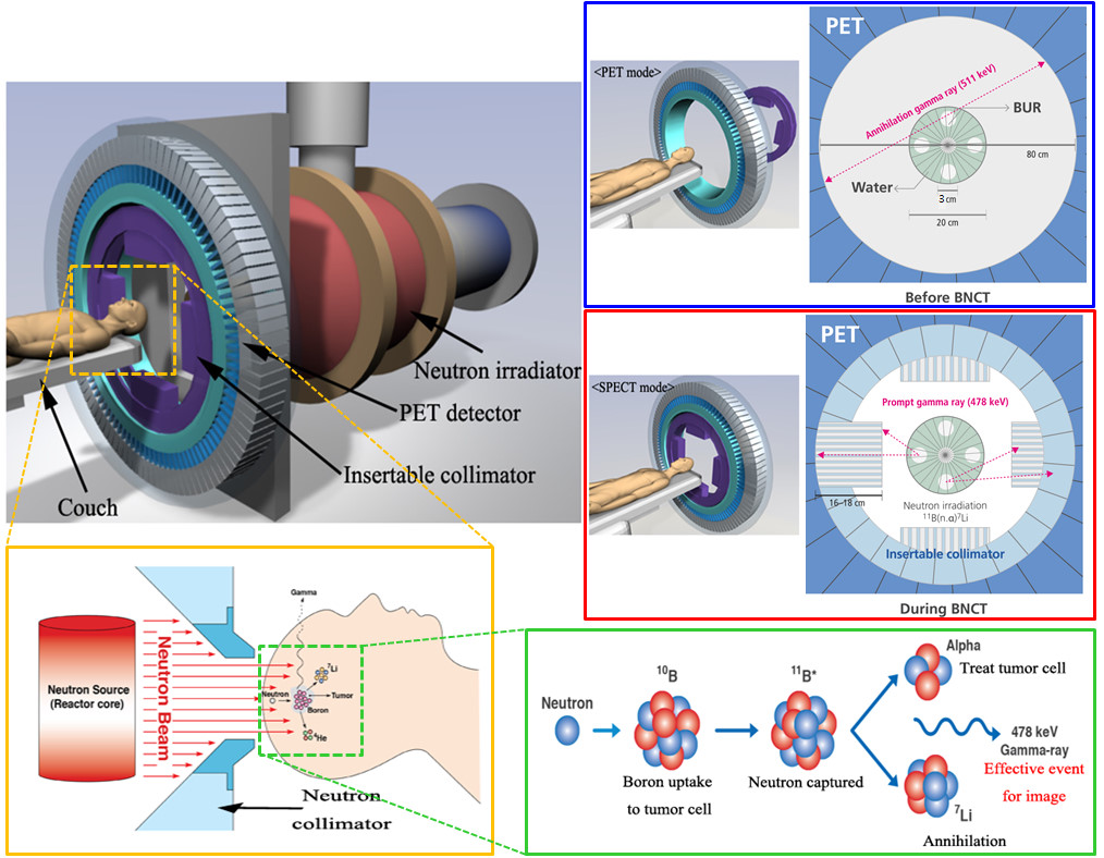
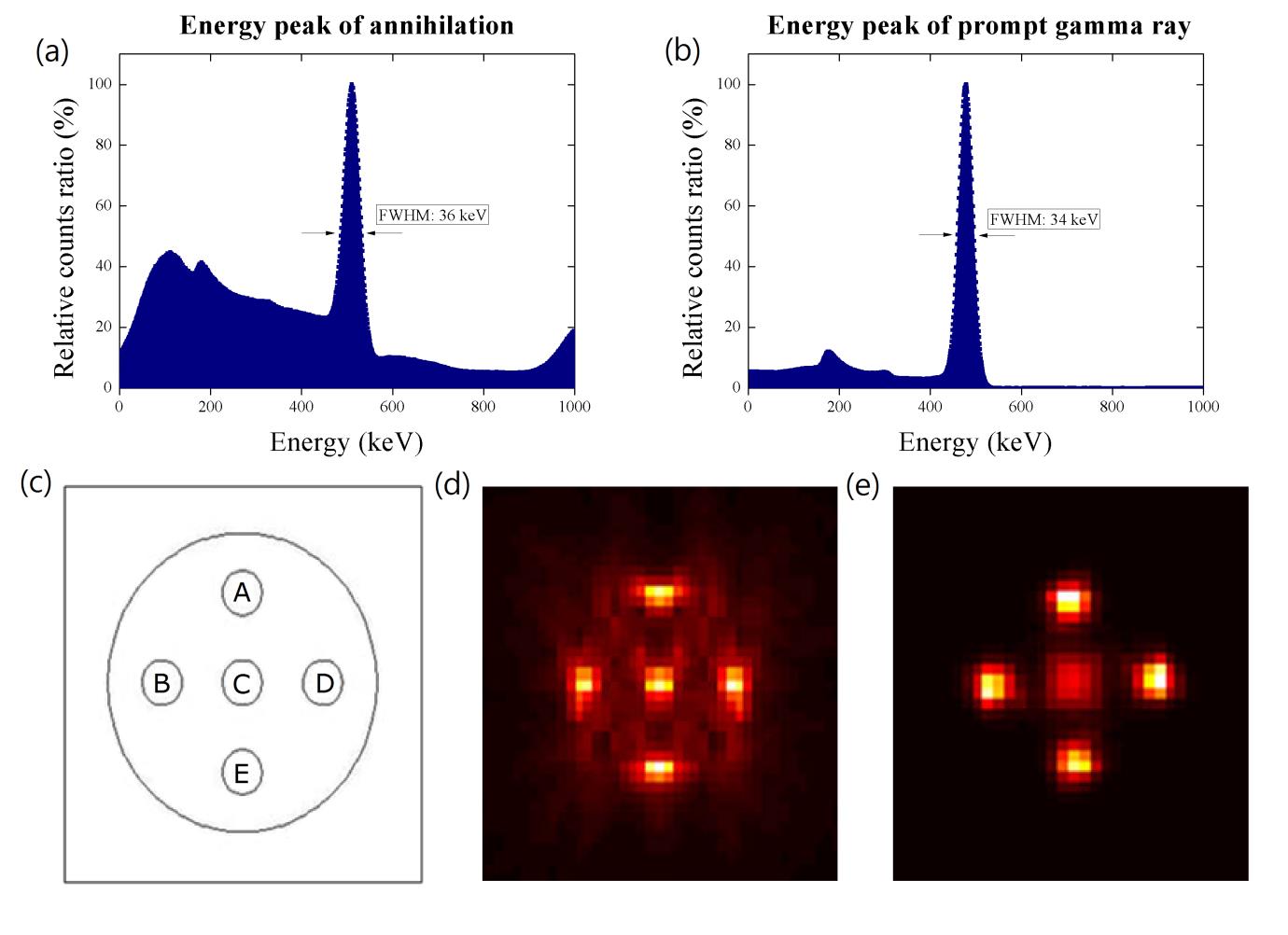
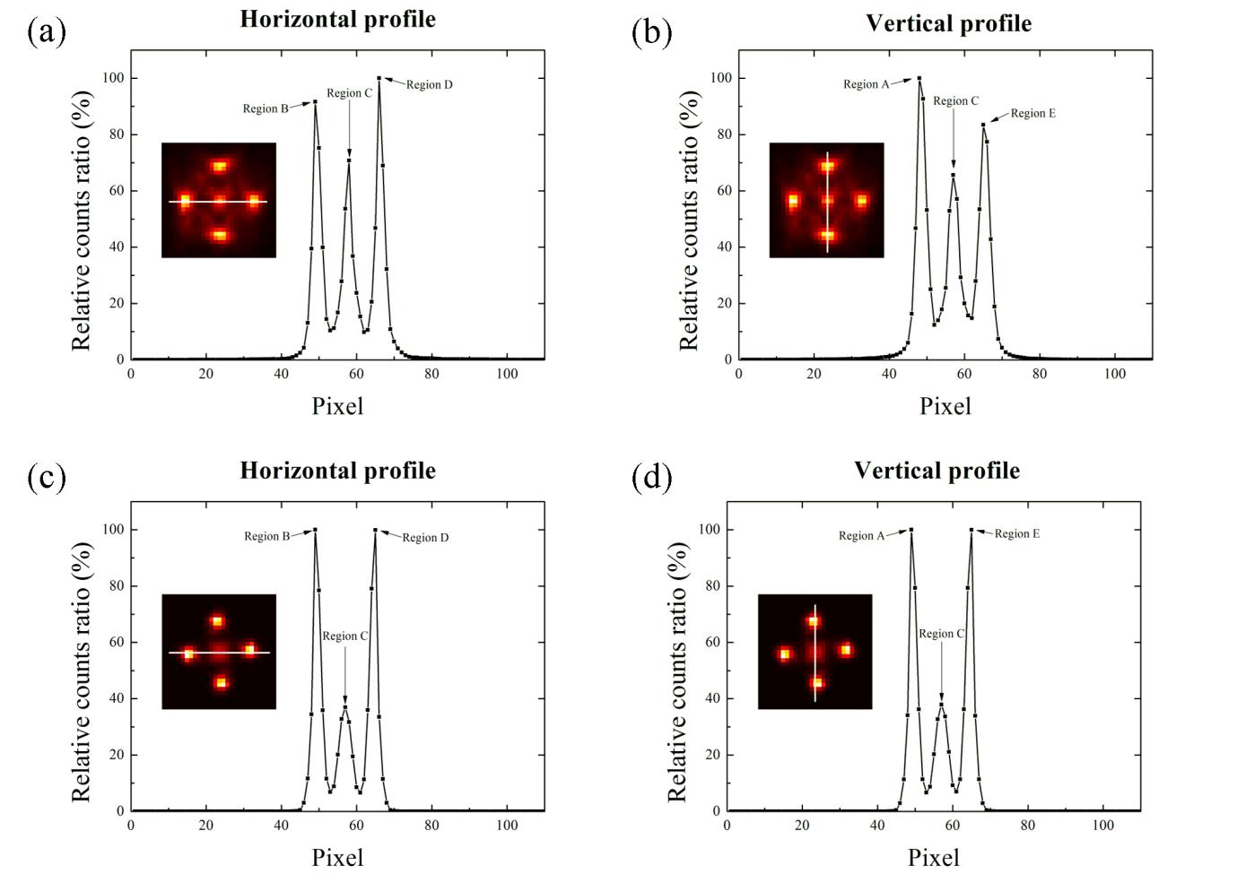
AbstractWe confirmed the feasibility of using our proposed system to extract two different kinds of functional images from a positron emission tomography (PET) module by using an insertable collimator during boron neutron capture therapy (BNCT). Coincidence events from a tumor region that included boron particles were identified by a PET scanner before BNCT; subsequently, the prompt gamma ray events from the same tumor region were collected after exposure to an external neutron beam through an insertable collimator on the PET detector. Five tumor regions that contained boron particles and were located in the water phantom and in the BNCT system with the PET module were simulated with Monte Carlo simulation code. The acquired images were quantitatively analyzed. Based on the receiver operating characteristic (ROC) curves in the five boron regions, A, B, C, D, and E, the PET and single-photon images were 10.2%, 11.7%, 8.2% (center region), 12.6%, and 10.5%, respectively. We were able to acquire simultaneously PET and single prompt photon images for tumor regions monitoring by using an insertable collimator without any additional isotopes.
Author information
Jung JY1, Lu B2, Yoon DK1, Hong KJ3, Jang H4, Liu C2, Suh TS5.
1Department of Biomedical Engineering, Research Institute of Biomedical Engineering, College of Medicine, Catholic University of Korea, Seoul 505, Republic of Korea.
2Department of Radiation Oncology, University of Florida, Gainesville, FL 32610-0385, United States.
3Molecular Imaging Program at Stanford (MIPS), Department of Radiology, Stanford University, 300 Pasteur Drive, Stanford, CA 94305, United States.
4Department of Radiation Oncology, College of Medicine, Seoul St. Mary's Hospital, Catholic University of Korea, Seoul 505, Republic of Korea.
5Department of Biomedical Engineering, Research Institute of Biomedical Engineering, College of Medicine, Catholic University of Korea, Seoul 505, Republic of Korea. Electronic address: suhsanta@catholic.ac.kr.
- 키워드
- Insertable collimator; PET; Prompt gamma ray; Tumor region
- 연구소개
- 붕소 중성자 포획 치료 (boron neutron capture therapy, BNCT)는 치료 전 종양 조직 내 붕소 표지화합물의 집적 정도를 PET으로 먼저 확인(annihilation gamma ray 포획)을 한 뒤에 (epi)thermal neuron을 조사하여 발생되는 alpha particle로 종양 조직을 치료하는 방법입니다. 이 때, 478 keV의 즉발 감마선 (prompt gamma ray)도 함께 방출이 되는 물리적 특성에 착안하여 본 연구에서는 PET에 삽입가능한 collimator를 탈/부착 하여 치료 중에 종양 조직을 실시간 모니터링 할 수 있는 방법을 제시하였습니다. 연구의 결과로 붕소가 집적된 종양 조직에서 재구성된 영상이 뚜렷하게 확인되었습니다. 본 연구의 장점은 치료 전과 치료 도중의 종양 변화를 기능적 영상을 이용하여 모니터링이 가능하다는 것입니다. 특히, 치료 도중에 획득한 영상으로 환자 셋업 및 처방 선량 오차 등을 사전에 확인하여 환자에게 불필요한 피폭을 막고, 정확하고 효과적인 치료를 제공할 수 있는 진단/치료 통합 시스템이라는 것이 본 연구의 특장점입니다. 현재 다시 각광받고 있는 BNCT에 대한 여러 제한점을 개선하고자 핵의학 장비의 장점을 활용한 융합 연구이며, 동시에 prompt gamma ray 방출의 의미를 부각시킨 연구로, 향후 방사선 치료 분야에서 붕소 표지 화합물을 접목시킨 새로운 연구도 활발히 진행될 것이라 사료됩니다.
- 덧글달기


