


글로벌 연구동향
의학물리학


![[Phys Med Biol .] TOPAS-imaging: extensions to the TOPAS simulation toolkit for medical imaging systems](/enewspaper/upimages/1686275542admin.PNG) [Phys Med Biol .] TOPAS-imaging: extensions to the TOPAS simulation toolkit for medical imaging systems
[Phys Med Biol .] TOPAS-imaging: extensions to the TOPAS simulation toolkit for medical imaging systems
Massachusetts General Hospital and Harvard Medical School / 이호연*
- 출처
- Phys Med Biol .
- 등재일
- 2023 Apr 3
- 저널이슈번호
- 68(8):10.1088/1361-6560/acc565. doi: 10.1088/1361-6560/acc565.
- 내용
Abstract
Objective. The TOol for PArticle Simulation (TOPAS) is a Geant4-based Monte Carlo software application that has been used for both research and clinical studies in medical physics. So far, most users of TOPAS have focused on radiotherapy-related studies, such as modeling radiation therapy delivery systems or patient dose calculation. Here, we present the first set of TOPAS extensions to make it easier for TOPAS users to model medical imaging systems.Approach. We used the extension system of TOPAS to implement pre-built, user-configurable geometry components such as detectors (e.g. flat-panel and multi-planar detectors) for various imaging modalities and pre-built, user-configurable scorers for medical imaging systems (e.g. digitizer chain).Main results. We developed a flexible set of extensions that can be adapted to solve research questions for a variety of imaging modalities. We then utilized these extensions to model specific examples of cone-beam CT (CBCT), positron emission tomography (PET), and prompt gamma (PG) systems. The first of these new geometry components, the FlatImager, was used to model example CBCT and PG systems. Detected signals were accumulated in each detector pixel to obtain the intensity of x-rays penetrating objects or prompt gammas from proton-nuclear interaction. The second of these new geometry components, the RingImager, was used to model an example PET system. Positron-electron annihilation signals were recorded in crystals of the RingImager and coincidences were detected. The simulated data were processed using corresponding post-processing algorithms for each modality and obtained results in good agreement with the expected true signals or experimental measurement.Significance. The newly developed extension is a first step to making it easier for TOPAS users to build and simulate medical imaging systems. Together with existing TOPAS tools, this extension can help integrate medical imaging systems with radiotherapy simulations for image-guided radiotherapy.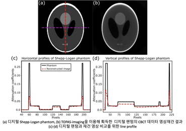
Affiliations
Hoyeon Lee 1, Bo-Wi Cheon 2, Joseph W Feld 3, Kira Grogg 4, Joseph Perl 5, José A Ramos-Méndez 6, Bruce Faddegon 6, Chul Hee Min 2, Harald Paganetti 1, Jan Schuemann 1
1Department of Radiation Oncology, Massachusetts General Hospital and Harvard Medical School, Boston, MA 02114, United States of America.
2Department of Radiation Convergence Engineering, Yonsei University, Wonju, Gangwon-do 26493, Republic of Korea.
3Electrical Engineering and Computer Science Department, Massachusetts Institute of Technology, Cambridge, MA 02139, United States of America.
4The Gordon Center for Medical Imaging, Department of Radiology, Massachusetts General Hospital and Harvard Medical School, Boston, MA 02114, United States of America.
5SLAC National Accelerator Laboratory, Menlo Park, CA 94025 United States of America.
6Department of Radiation Oncology, University of California San Francisco, San Francisco, CA 94115 United States of America.
- 키워드
- Monte carlo; cone-beam CT; medical imaging; positron emission tomography; prompt gamma.
- 연구소개
- 본 연구에서는 방사선 치료의 전산모사를 위해서 활발히 이용되고 있는 Monte Carlo 코드 중 하나인 TOols for PArticle Simulation (TOPAS)를 이용해 의료영상 기기 전산모사를 위한 extension을 개발하였습니다. 개발한 extension은 질병의 진단과 방사선 치료의 정확도 향상을 위해서 이용되고 있는 Cone-Beam Computed Tomography (CBCT), Positron Emission Tomography (PET), Single Photon Emission Computed Tomography (SPECT), 그리고 Prompt Gamma 시스템을 모델링하기 위해서 검출기, collimator 등의 하드웨어와 의료영상기기를 통해서 획득한 신호처리를 위한 scorer를 지원합니다. 본 연구에서 개발한 extension의 시연을 위해서 디지털 팬텀을 구성하고 CBCT와 PET 시스템을 모델링하여 영상을 획득하였고, 영상재건 소프트웨어를 이용하여 3차원 영상을 획득하였습니다. 또한 양성자의 range verification 에 이용되는 Prompt gamma 시스템을 모델링하였습니다. 개발한 코드는 모두 TOPAS 와 함께 Open-source로 제공될 예정으로 의료영상 기기 관련 연구를 수행하는 기관에서 자유롭게 이용하실 수 있습니다.
- 덧글달기


![]()
- COPYRIGHT(C) 2015 한국원자력의학원 전략기획팀 All rights Reserved.
- 문의 : rmwebzine@kirams.re.kr 발행처 : 한국원자력의학원 전략기획팀
- 우) 01812 서울시 노원구 노원로 75 한국원자력의학원 전략기획팀
편집위원
몬테카를로 시뮬레이션을 통해 방사선치료분야에서 선량 등을 계산하는 연구는 많이 시행되어 왔으나 영상시스템을 모사하는 연구는 생소하다. 방사선치료 및 영상의학분야에 많은 도움이 되었으면 한다.
2023-06-09 09:20:42