
글로벌 연구동향
핵의학


![[Sci Rep.] The role of 18 F-fluorodeoxyglucose-positron emission tomography/computed tomography in the differential diagnosis of pericardial disease](/enewspaper/upimages/1612332165admin.JPG) 2021년 02월호
2021년 02월호
[Sci Rep.] The role of 18 F-fluorodeoxyglucose-positron emission tomography/computed tomography in the differential diagnosis of pericardial disease성균관의대 / 현철원, 이현경, 최준영*, 장성아*
- 출처
- Sci Rep.
- 등재일
- 2020 Dec 9
- 저널이슈번호
- 10(1):21524. doi: 10.1038/s41598-020-78581-y.
- 내용
Abstract
This study aimed to assess the role of 18F-fluorodeoxyglucose-positron emission tomography/computed tomography (18FDG-PET/CT) in the differential diagnosis of pericardial disease. The diagnosis is often troublesome because pericardial fluid analysis or biopsy does not always provide answers. 18FDG-PET/CT can visualize both inflammation and malignancy and offers a whole-body assessment. Patients who visited the Pericardial Disease Clinic of Samsung Medical Center with an 18FDG-PET/CT order code were extracted. Exclusion criteria were as follows: (1) the purpose of the differential diagnosis was not pericardial disease; (2) the patient had a known advanced-stage malignancy; (3) the patient already have confirmative diagnosis using a serology, pericardial effusion analysis or biopsy. The analysis included 107 patients. The most common final diagnosis was idiopathic (n = 46, 43.0%), followed by tuberculosis (n = 30, 28.0%) and neoplastic (n = 11, 10.3%). A maximum standardized uptake value (SUVmax) ≥ 5 typically indicates tuberculosis or neoplastic pericarditis except in just one case of autoimmune pericarditis); especially all of the SUVmax scores ≥ 10 had tuberculosis. The diagnostic yield of pericardial biopsy was very low (10.2%). Interestingly, all of the pericardium with an SUVmax < 4.4 had nondiagnostic results. In contrast, targeted biopsies based on 18FDG uptake demonstrated a higher diagnostic yield (38.7%) than pericardium. The sensitivity of 18FDG-PET/CT was 63.6%. The specificity was 71.9%. The positive predictive value was 20.6%. The negative predictive value 94.5%, and the accuracy was 71.0% for excluding malignancy based upon the FDG uptake patterns. It is possible to explore the differential diagnosis in some patients with difficult pericardiocentesis or pericardial biopsy in a noninvasive manner using on the SUVmax or uptake patterns. In addition, the biopsy strategy depending on 18FDG uptake is helpful to achieve biopsy more safely and with a higher yield. 18FDG-PET may enhance the diagnostic efficacy in patients with pericardial disease.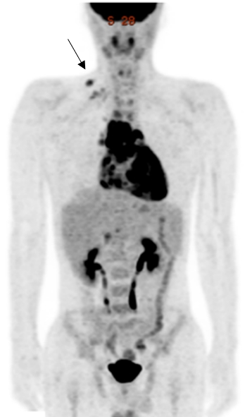
그림 설명: 심낭질환이 의심되는 환자에서 어려운 심낭생검 대신 PET/CT에서 보이는 우측 쇄골상림프절(검정 화살표)을 조직 검사하여 결핵성 심낭염으로 진단된 증례임.
Affiliations
Cheol Won Hyeon 1 , Hyun Kyung Yi 2 , Eun Kyoung Kim 1 3 , Sung-Ji Park 1 3 , Sang-Chol Lee 1 3 , Seung Woo Park 1 3 , Jae K Oh 4 , Joon Young Choi 5 , Sung-A Chang 6 7
1 Division of Cardiology, Department of Medicine, Samsung Medical Center, Sungkyunkwan University School of Medicine, 81 Irwon-ro, Gangnam-gu, Seoul, 06351, Republic of Korea.
2 Department of Nuclear Medicine, Samsung Medical Center, Sungkyunkwan University School of Medicine, 81 Irwon-ro, Gangnam-gu, Seoul, 06351, Republic of Korea.
3 Heart Vascular Stroke Imaging Center, Heart Vascular Stroke Institute, Samsung Medical Center, Sungkyunkwan University School of Medicine, Seoul, Republic of Korea.
4 Department of Cardiovascular Medicine, Mayo Clinic College of Medicine, Rochester, USA.
5 Department of Nuclear Medicine, Samsung Medical Center, Sungkyunkwan University School of Medicine, 81 Irwon-ro, Gangnam-gu, Seoul, 06351, Republic of Korea. jynm.choi@samsung.com.
6 Division of Cardiology, Department of Medicine, Samsung Medical Center, Sungkyunkwan University School of Medicine, 81 Irwon-ro, Gangnam-gu, Seoul, 06351, Republic of Korea. elisabet.chang@gmail.com.
7 Heart Vascular Stroke Imaging Center, Heart Vascular Stroke Institute, Samsung Medical Center, Sungkyunkwan University School of Medicine, Seoul, Republic of Korea. elisabet.chang@gmail.com.
- 연구소개
- 다양한 종류의 심낭질환들의 FDG PET/CT 소견을 비교 분석함으로써 이들 질환의 감별진단에 FDG PET/CT가 도움을 줄 수 있다는 것을 보여준 임상적으로 의미 있는 연구입니다. 특히. 조직학적 검사가 위음성이 나오기 쉬운 통상적인 심낭생검 대신 시 PET/CT 유도 생검이 진단율 향상에 도움을 줄 수 있다는 것도 보여주었습니다.
- 덧글달기


