
글로벌 연구동향
핵의학


![[J Vasc Interv Radiol.] Chitosan-Based Hydrogel Microparticles for Treatment of Carcinoma in a Rabbit VX2 Liver Tumor Model.](/enewspaper/upimages/1524118944admin.PNG) 2018년 04월호
2018년 04월호
[J Vasc Interv Radiol.] Chitosan-Based Hydrogel Microparticles for Treatment of Carcinoma in a Rabbit VX2 Liver Tumor Model.전북의대 / 황효숙, 김현수, 정환정*
- 출처
- J Vasc Interv Radiol.
- 등재일
- 2018 Apr
- 저널이슈번호
- 29(4):575-583. doi: 10.1016/j.jvir.2017.11.032. Epub 2018 Feb 22.
- 내용
Abstract
PURPOSE:
To investigate potential of chitosan hydrogel microparticles (CHI) for treatment of VX2carcinoma.MATERIALS AND METHODS:
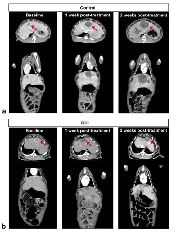
Two weeks after liver VX2 implantation, contrast-enhanced computerized tomographic scanning was conducted. Rabbits (n = 2) with successful tumor growth were treated with different sizes of 99mTc-labeled CHI (60-80 μm and 100-120 μm) via intra-arterial hepatic catheterization. Liver distribution of 99mTc-labeled CHI was determined by means of autoradiography, a radiation-based photographic technique. In the next part of this study, therapeutic effectiveness was examined with the use of CHI with the size range of 60-80 μm (n = 11). Tumor growth response and levels of blood liver enzymes were studied at baseline and 1 and 2 weeks after CHI treatment.RESULTS:
Successful tumor growth was confirmed in all rabbits (24/24). Intrahepatic CHI with the size range of 60-80 μm resulted in liver localization in more close proximity to tumor nodule versus 100-120 μm. Baseline tumor volume was 1,909 ± 575 mm3 in animals receiving CHI versus 1,831 ± 249 mm3 in control animals (P = .342). In control animals, tumor volume markedly increased by 1,544 ± 512% at 2 weeks after sham operation versus baseline. In animals receiving CHI, tumorvolume remained relatively unchanged (54 ± 6% increase; P = .007 vs control). Levels of blood aspartate transaminase (AST) and alanine transaminase (ALT) in animals receiving CHI increased 1 week after treatment (P = .032 vs control for AST; P = .000 vs control for ALT), but returned to control levels at 2 weeks.CONCLUSIONS:
CHI embolization suppressed tumor growth without appreciable damages in liverfunction.Author information
Hwang H1, Kim HS1, Kwon J1, Oh PS1, Park HS2, Lim ST1, Sohn MH1, Jeong HJ3.
1 Department of Nuclear Medicine, Molecular Imaging and Therapeutic Medicine Research Center, Cyclotron Research Center, Research Institute of Clinical Medicine, Biomedical Research Institute, Chonbuk National University Medical School and Hospital, 634-18 GeumAm-dong, Duckjin-gu, Jeonju-si, Jeollabuk-do 561-803, Republic of Korea.
2 Department of Pathology, Chonbuk National University Medical School and Hospital, Jeonju, Jeonbuk, Republic of Korea.
3 Department of Nuclear Medicine, Molecular Imaging and Therapeutic Medicine Research Center, Cyclotron Research Center, Research Institute of Clinical Medicine, Biomedical Research Institute, Chonbuk National University Medical School and Hospital, 634-18 GeumAm-dong, Duckjin-gu, Jeonju-si, Jeollabuk-do 561-803, Republic of Korea. Electronic address: jayjeong@jbnu.ac.kr.
- 연구소개
- 키토산을 이용한 하이드로겔을 제조하여 간암 토끼 모델에서의 치료효과를 입증한 논문입니다. 색전술제재를 개발하기 위해서 결정되어야 할 사항들은 입자 제조법 및 안정성, 색전술제재의 입자의 크기에 따른 암조직에서의 분포의 차이, 색전술을 통한 치료효과 규명 등입니다. 연구방법적 측면에서 VX2 모델의 안정적 구축 및 유지기술, 조영 증강 CT영상 촬영법도 확보, 대퇴동맥을 통한 마이크로카테터를 삽입술 및 간동맥 초선택적 색전치료술 등이 활용되었습니다. 개발된 하이드로겔은 종양의 성장부위인 가장자리쪽으로 잘 분포됨으로써 좋은 간암치료효과를 보이는 것을 확인하였습니다.
- 덧글달기


편집위원
Chitosan hydrogel microparticle을 개발하고 이를 이용하여 핵의학 치료영역을 넓힌 연구로 핵의학 영역확장에 좋은 영향을 미칠 수 있는 연구임. 기초연구자 뿐만 아니라 종양관련 의사와 임상 핵의학자에게도 관심을 끌 논문으로 생각됨.
덧글달기닫기2018-04-17 10:58:59
등록