글로벌 연구동향
핵의학
![[Clin Nucl Med.] 18F-FDG PET/CT Versus Contrast-Enhanced CT for Staging and Prognostic Prediction in Patients With Salivary Gland Carcinomas.](/enewspaper/upimages/admin_20170413112559_R.png) [Clin Nucl Med.] 18F-FDG PET/CT Versus Contrast-Enhanced CT for Staging and Prognostic Prediction in Patients With Salivary Gland Carcinomas.
[Clin Nucl Med.] 18F-FDG PET/CT Versus Contrast-Enhanced CT for Staging and Prognostic Prediction in Patients With Salivary Gland Carcinomas.울산의대 / 박만준, 노종렬*
- 출처
- Clin Nucl Med.
- 등재일
- 2017 Mar
- 저널이슈번호
- 42(3):e149-e156. doi: 10.1097/RLU.0000000000001515.
- 내용

FIGURE 1. Preoperative detection of metastatic lymph nodes in the neck. In a 54-year-old woman with salivary duct carcinoma (arrow, A and B) on the right submandibular gland, 18F-FDG PET/CT shows a metastatic lymph node in the neck (arrowheads, B and C) that was not properly identified by contrast-enhanced CT (D).
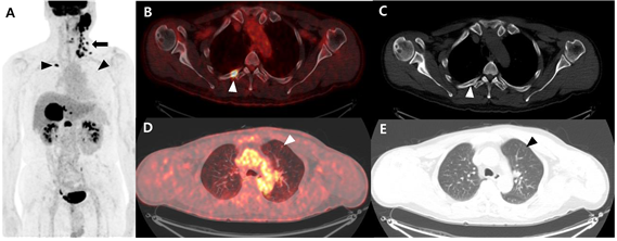
FIGURE 2. Pretreatment detection of distant metastasis. In a 58-year-old man with salivary duct carcinoma, 18F-FDG PET/CT shows the metastatic diseases in the neck (arrow, A) and the rib and lung (arrowheads, B and C) that were not all detected by CT scans (D and E).
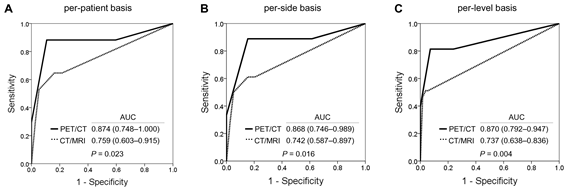
FIGURE 3. Receiver operating curve analyses comparing the diagnostic values of 18F-FDG PET/CT and contrast-enhanced CT for detection of cervical nodal metastases of SGC according to per-patient analysis. (A), per-neck side analysis (B), and per-cervical level analyses (C). The AUCs were separately calculated, and the statistical differences of AUC values between 18F-FDG PET/CT and CT were then compared (P < 0.05).
Abstract
PURPOSE:
Salivary gland carcinoma (SGC) is rare tumor with various histological type and metastatic potential. Pretreatment detection of metastases can contribute to planning the appropriate treatment of SGC. Therefore, the present study evaluated the utility of F-FDG PET/CT versus contrast-enhanced CT for detection of metastases and prediction of outcomes in SGC patients.
METHODS:
Sixty-seven consecutive SGC patients who were prospectively evaluated by F-FDG PET/CT and contrast-enhanced CT and subsequently underwent surgery with or without postoperative radiotherapy/chemoradiotherapy were included. The diagnostic values of both imaging modalities for detection of metastatic diseases were compared with McNemar test and logistic regression using generalized estimating equations. Cox proportional hazard modeling was used to assess the prognostic values of the quantitative metabolic measurements detected by F-FDG PET/CT and of other clinical factors.
RESULTS:
Among 67 SGC patients, 17 (25.4%) had cervical metastasis, and 4 (6%) had distant metastasis at initial staging. The sensitivity of F-FDG PET/CT for detection of cervical metastasis was significantly higher than those of CT (P < 0.05), and those of F-FDG PET/CT and CT for detection of distant metastasis did not differ (P > 0.5). Regional and distant site metastases were most reliably predicted by high-grade pathological analysis (P < 0.05). Extranodal extension and metabolic tumor volume measured by F-FDG PET/CT were independent predictors of progression-free survival and overall survival (all P < 0.05).
CONCLUSIONS:
In SGC patients, F-FDG PET/CT detected metastatic diseases with high sensitivity and specificity, and metabolic tumor volumes helped to predict survival outcomes.
Author information
Park MJ1, Oh JS, Roh JL, Kim JS, Lee JH, Nam SY, Kim SY.
1 From the Departments of *Otolaryngology, †Nuclear Medicine, and ‡Radiology, Asan Medical Center, University of Ulsan College of Medicine, Seoul, Republic of Korea.
- 연구소개
- 타액선, 흔히 침샘으로 불리는 타액선에 발생하는 암종은 드물게 발생하며 그 종류도 매우 다양한 암종입니다. 타액선에 발생하는 암종의 진단함에 있어, 치료 계획을 확립하기 위하여 정밀한 병변이 범위 및 병기 설정은 무엇보다 중요합니다. 본 연구진은 18F-FDG PET/CT 가 기존에 통상적으로 사용된 진단도구인 경부 및 흉부의 조영증강 CT 에 비하여 더 우수한 진단적 효용성을 보이는지를 본 연구를 통하여 밝혀내고자 하였습니다. 또한, 얻어진 PET 영상에서 해당 암종이 보이는 위치의 평균 signal 을 측정하여 SUVmean 값을 구하였으며, PET 영상에서 그려지는 종양의 부피인 metabolic tumor volume (MTV)를 계산하였습니다. Total total lesion glycolysis (TLG) 는 SUVmean과 MTV를 곱하여 나온 값으로, 해당 병변의 포도당 대사 정도를 반영하며, 종양의 생리학적/기능적 burden을 대표하는 임상 표지자로서, 많은 기존의 암에서 예후인자로서 그 유용성이 확인된 바 있습니다. 본 연구에서는, MTV, TLG와 같은 18F-FDG PET으로부터 유래된 지표들이 타액선암의 예후과 관련이 있는지를 또한 보고자 하였습니다. 본 연구는 타액선 암종으로 새로이 진단된 환자를 전향적으로 선별하여 시행되었으며, 총 67명의 환자가 연구대상으로 선별되었습니다. 전체 환자에 있어 18F-FDG PET/CT는 기존의 조영증강 CT 에 비하여 원격 전이의 발견율의 차이는 보이지 않았으나 (P>0.05), 경부 림프절 전이의 진단에 있어 더 높은 민감도 및 특이도를 보였습니다 (P<0.05). 또한, MTV값이 높을수록, 높은 재발율 및 사망률을 예측하는 독립적인 위험인자가 됨을 발견하였습니다 (P<0.05). 본 연구는 타액선 악성 종양의 진단 및 병기 설정에 있어, 기존의 CT 보다 18F-FDG PET/CT가 더 우수함을 보여주며, 18F-FDG PET/CT로부터 계산될 수 있는 생리학적 종양지표들은 환자의 예후를 예측할 수 있는 중요한 임상 표지자가 됨을 보고합니다.
- 덧글달기


