글로벌 연구동향
핵의학
![[J Nucl Med.] Novel PET Imaging of Atherosclerosis with 68Ga-Labeled NOTA-Neomannosylated Human Serum Albumin](/enewspaper/upimages/admin_20161214135916_R.jpg) 2016년 12월호
2016년 12월호
[J Nucl Med.] Novel PET Imaging of Atherosclerosis with 68Ga-Labeled NOTA-Neomannosylated Human Serum Albumin서울의대 / 김응주,김성은,서홍석*, 정재민*
- 출처
- J Nucl Med.
- 등재일
- 2016 Nov
- 저널이슈번호
- 57(11):1792-1797. Epub 2016 Jun 23.
- 내용
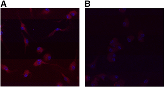
그림1. Mannose 수용체에 결합하는 RITC-mannosylated human albumin(MSA). A, C57/BL6 생쥐의 복막 거식세포들 배양에서 RITC-MSA 투여후 confocal 현미경으로 세포에 결합된 RITC-MSA가 관찰됨. B, 같은 조건에서 anti-CD206 MR blocking 항체처리후 RITC-MSA를 투여 배양하면 세포에 RITC-MSA가 거의 결합되지 않음.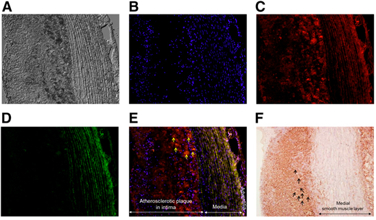
그림2. 콜레스테롤 식이를 이용한 토끼의 대동맥부위 죽상동맥경화부위에 선택적으로 결합하는 RITC-MSA의 현미경 영상. A, 죽상동맥경화병소의 DIC(Differential interference contrast)현미경 영상. B, 같은 병소부위의 DAPI-염색한 세포핵 영상. C, 거식세포에 결합하여 죽상경화반 병소에 광범위하게 분포한 RITC-MSA의 영상. D, MSA가 포함되지 않은 FITC 영상으로 죽상경화부위에 거의 결합되지 않음. E, B-E의 결합영상으로 화살표부위의 죽상경화부위의 거식세포에 결합된 RITC-MSA의 분자영상. F, 근처 대동맥부위의 죽상동맥 경화병소에 존재하는 거식세포들의 면역화학염색 영상(화살표부위의 거식세포들)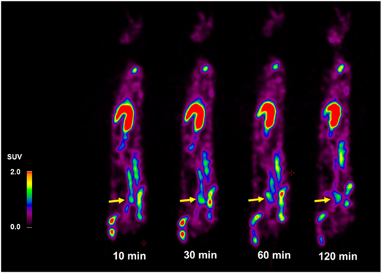
그림3. 대동맥 부위의 죽상경화 68Ga-NOTA-MSA을 이용한 PET 영상으로 주사후 10-120분사이에 촬영한 바 죽상경화 부위(화살표 표시부위)의 영상이 in vivo상태에서 maximal SUV가 1.7, 1.8, 1.5, 1.2로 유지되었음.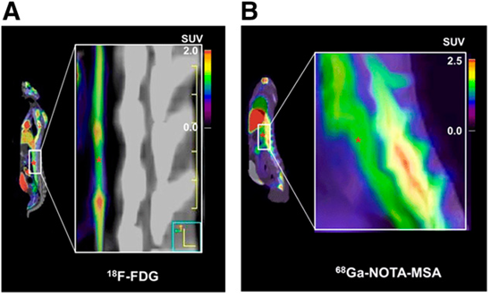
그림4. 대동맥 죽상경화병소에서 18F-FDG PET영상과 68Ga-NOTA-MSA PET영상의 비교. A, 18F-FDG 영상으로 확대된 영상에서 초록색(붉은색 별표시) 부위의 죽상경화부위에 영상이 관찰됨. B, 68Ga-NOTA-MSA 영상으로 확대된 영상에서 역시 초록색(붉은색 별표시) 부위의 죽상경화부위에 영상이 잘 관찰됨.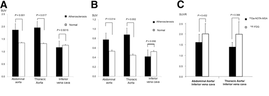
그림5. 대동맥 죽상경화병소에서 68Ga-NOTA-MSA PET영상과 18F-FDG PET영상에서 측정한 SUV값의 비교. A, 68Ga-NOTA-MSA PET 영상에서 복부대동맥, 흉부대동맥, 대정맥 부위의 SUV측정값의 죽상경화동물과 정상동물의 비교한바 죽상경화 병소부위에서 유의한 차이가 남. B,18F-FDG PET 영상에서 복부대동맥, 흉부대동맥, 대정맥 부위의 SUV측정값의 죽상경화동물과 정상동물의 비교한바 병소부위에서 차이가 남. C, 68Ga-NOTA-MSA PET영상과 18F-FDG PET영상에서 측정한 SUV값을 각각의 대정맥 SUV값으로 나눈 비 값은 68Ga-NOTA-MSA PET영상과 18F-FDG PET영상에서 차이가 없기 때문에 같은 정도의 영상 차이를 얻었다고 판단됨.Abstract
Activated macrophages take up 18F-FDG via glucose transporters, so this compound is useful for atherosclerosis imaging by PET. However, 18F-FDG application is limited for imaging of the heart and brain, in which glucose uptake is high, and in patients with aberrant glucose metabolism. The aims of this study were to confirm that mannosylated human serum albumin (MSA) specifically binds to the mannose receptor (MR) on macrophages and to test the feasibility of 68Ga-labeled NOTA-MSA for PET imaging of atherosclerotic plaques.
METHODS:
The peritoneal macrophages of C57/B6 mice were collected, incubated with rhodamine B isothiocyanate-MSA (10 μg/mL), and evaluated by confocal microscopy and flow cytometry. The same evaluations were performed after preincubation of the macrophages with anti-CD206 MR blocking antibodies. NOTA-MSA was synthesized by conjugating 2-(p-isothiocyanatobenzyl)-1,4,7-triazacyclononane-1,4,7-triacetic acid to MSA, followed by labeling with 68Ga. Rabbits with atherosclerotic aorta induced by a 3-mo cholesterol diet and chronic inflammation underwent consecutive PET/CT with 18F-FDG and 68Ga-NOTA-MSA at 2-d intervals.
RESULTS:
The binding of MSA to MR and its dose-dependent reduction by preincubation with anti-CD206 MR blocking antibody were confirmed. Rhodamine B isothiocyanate and fluorescein isothiocyanate fluorescence colocalized at the atherosclerotic plaque. The 68Ga-NOTA-MSA SUVs of the atherosclerotic aorta were significantly higher than those of the healthy arteries and inferior vena cava and were comparable to those obtained with 18F-FDG.
CONCLUSION:
These findings suggest that MR-specific 68Ga-NOTA-MSA is effective for detecting atherosclerosis in the aorta and is a promising radiopharmaceutical for imaging atherosclerosis because of the presence of M2 macrophages in atherosclerotic plaques.
Author information
Kim EJ1, Kim S2, Seo HS3,4, Lee YJ1, Eo JS2, Jeong JM5, Lee B6, Kim JY7, Park YM8, Jeong M8.
1Cardiovascular Center, Korea University Guro Hospital, Korea University College of Medicine, Seoul, South Korea.
2Nuclear Medicine, Korea University Guro Hospital, Korea University College of Medicine, Seoul, South Korea.
3Cardiovascular Center, Korea University Guro Hospital, Korea University College of Medicine, Seoul, South Korea mdhsseo@korea.ac.kr jmjng@snu.ac.kr.
4Korea University-Korea Institute of Science and Technology (KU-KIST) Graduate School of Converging Science and Technology, Seoul, South Korea.
5Department of Nuclear Medicine, Institute of Radiation Medicine, Seoul National University College of Medicine, Seoul, South Korea mdhsseo@korea.ac.kr jmjng@snu.ac.kr.
6Department of Nuclear Medicine, Institute of Radiation Medicine, Seoul National University College of Medicine, Seoul, South Korea.
7Research Institute of Skin Imaging, Korea University College of Medicine, Seoul, South Korea; and.
8Department of Molecular Medicine, Ewha Womans University School of Medicine, Seoul, South Korea.
- 키워드
- atherosclerosis; inflammation; mannose receptor; positron emission tomography
- 연구소개
- 본 연구는 심혈관질환의 가장 중요한 기저 원인으로 알려진 죽상동맥경화의 분자영상을 거식세포의 mannose 수용체에 결합하는 mannosylated human albumin(MSA) 에 chelating agent인 NOTA를 결합하여 68Ga을 표지시켜 PET을 이용하여 비관혈적인 방법으로 in vivo 상태로 구현한 연구이다. 본 연구에 사용한 68Ga-NOTA-MSA는 서울대학교 핵의학과 정재민 교수가 간에서 Kupffer cell 분자 영상용 물질로 독자적으로 개발한 바 있으며, 이를 죽상동맥 경화 분자영상 구현에 응용하여 성공한 연구이다. 아직 임상적으로 사용가능한 죽상동맥경화용 PET probe는 없는 상태이지만 연구용으로 일부 사용하는 18F-FDG(fluorinated deoxyglucose) 와 18F-FDM(fluorinated deoxymannose)의 경우는 죽상동맥경화 병소를 이루는 주요세포인 거식세포의 대사적 특성을 이용하기 때문에 뇌와 심장의 경우에서 적용이 어렵고, 또한 당뇨 등의 대사적 장애가 있는 경우에도 사용이 제한적이나, 68Ga-NOTA-MSA는 거식세포의 세포막에 존재하는 수용체의 특성을 이용하기 때문에 대사적 특성의 사용한계가 문제되지 않아 임상적 적용에 특별한 대사적 제한이 없는 것이 특징이다. 또한 양전자 방출핵종인 68Ga는 제너레이터에서 쉽게 얻을 수 있어 사이클로트론과 같은 비싸고 복잡한 장비가 있어야 하는 18F로 표지된 방사성의약품에 비하여 임상 적용이 훨씬 더 편리하다는 장점도 있다.
- 덧글달기


편집위원
핵의학분자영상 기법을 이용한 동맥경화 검색에 대한 연구 논문으로 기초연구에 관심이 있는 임상핵연구자나 방사화학관련 연구자들에게 관심을 끌 논문으로 보입니다.
덧글달기닫기2016-12-06 09:31:40
등록
편집위원2
FDG만으로 한계를 느끼는 요즈음 종양분야가 아닌 염증 및 기타 양성질환 분야의 tacer 개발 또는 임상적 유용성 증명은 의미가 있다고 생각합니다.
2016-12-06 09:31:40