


글로벌 연구동향
핵의학


![[Ann Nucl Med .] Quantitative comparative analysis of amyloid PET images using three radiopharmaceuticals](/enewspaper/upimages/1688963926admin.PNG) [Ann Nucl Med .] Quantitative comparative analysis of amyloid PET images using three radiopharmaceuticals
[Ann Nucl Med .] Quantitative comparative analysis of amyloid PET images using three radiopharmaceuticals
동아의대 / 정영진*
- 출처
- Ann Nucl Med .
- 등재일
- 2023 May
- 저널이슈번호
- 37(5):271-279. doi: 10.1007/s12149-023-01824-1. Epub 2023 Feb 7.
- 내용
Abstract
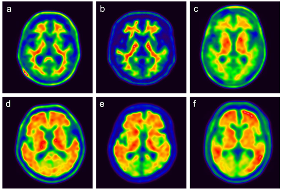
Objective: Amyloid positron emission tomography (PET) with F-18 florbetaben (FBB), F-18 flutemetamol (FMM), and F-18 florapronol (FPN) is being used clinically for the evaluation of dementia. These radiopharmaceuticals are commonly used to evaluate the accumulation of beta-amyloid plaques in the brain, but there are structural differences between them. We investigated whether there are any differences in the imaging characteristics.Methods: A total of 605 subjects were enrolled retrospectively in this study, including healthy subjects (HS) and patients with mild cognitive impairment or Alzheimer's disease. Participants underwent amyloid PET imaging using one of the three radiopharmaceuticals. The PET images were analyzed visually and semi-quantitatively using a standardized uptake value ratio (SUVR). In addition, we calculated and compared the cut-off SUVR of the representative regions for each radiopharmaceutical that can distinguish between positive and negative scans.
Results: In the negative images of the HS group, the contrast between the white matter and the gray matter was high in the FMM PET images, while striatal uptake was relatively higher in the FPN PET images. The SUVR showed significant differences across the radiopharmaceuticals in all areas except the temporal lobe, but the range of differences was relatively small. Accuracy levels for the global cut-off SUVR to discriminate between positive and negative images were highest in FMM PET, with a value of 0.989. FBB PET also showed a high value of 0.978, while FPN PET showed a relatively low value of 0.901.
Conclusions: Negative amyloid PET images using the three radiopharmaceuticals showed visually and quantitatively similar imaging characteristics except in the striatum. Binary classification using the cut-off of the global cortex showed high accuracy overall, although there were some differences between the three PET images.
Affiliations
Young Jin Jeong 1 2, Hyun Jin Yoon 3, Do-Young Kang 3 4 5, Kyung Won Park 6
1Department of Nuclear Medicine, Dong-A University Hospital, Dong-A University College of Medicine and Medical Center, 1, 3ga, Dongdaesin-dong, Seo-gu, Busan, 602-715, Republic of Korea. nmjeong@gmail.com.
2Institute of Convergence Bio-Health, Dong-A University, Busan, Republic of Korea. nmjeong@gmail.com.
3Department of Nuclear Medicine, Dong-A University Hospital, Dong-A University College of Medicine and Medical Center, 1, 3ga, Dongdaesin-dong, Seo-gu, Busan, 602-715, Republic of Korea.
4Institute of Convergence Bio-Health, Dong-A University, Busan, Republic of Korea.
5Department of Translational Biomedical Sciences, Dong-A University, Busan, Republic of Korea.
6Department of Neurology, Dong-A University Hospital, Dong-A University College of Medicine, Busan, Republic of Korea.
- 키워드
- Amyloid PET; Cut-off; F-18 florapronol; F-18 florbetaben; F-18 flutemetamol; Standardized uptake value ratio.
- 연구소개
- Amyloid PET 검사를 위해 국내에서 사용되고 있는 3종의 방사성의약품 (F-18 florbetaben, F-18 flutemetamol, F-18 florapronol)의 음성과 양성 영상을 육안 및 정량적으로 비교한 연구입니다. 모든 검사는 동일한 병원에서 동일한 기기를 이용하여 얻었기 때문에 검사 프로토콜과 기기의 차이로 인한 영상의 차이를 최대한 배제할 수 있었습니다. 단일 기관에서 얻은 3가지 PET 영상의 특성을 비교해 볼 수 있는 좋은 정보라고 생각됩니다.
- 덧글달기


![]()
- COPYRIGHT(C) 2015 한국원자력의학원 전략기획팀 All rights Reserved.
- 문의 : rmwebzine@kirams.re.kr 발행처 : 한국원자력의학원 전략기획팀
- 우) 01812 서울시 노원구 노원로 75 한국원자력의학원 전략기획팀