
글로벌 연구동향
핵의학


![[Int J Mol Sci.] Identification of Lymphatic and Hematogenous Routes of Rapidly Labeled Radioactive and Fluorescent Exosomes through Highly Sensitive Multimodal Imaging](/enewspaper/upimages/1606889373admin.JPG) 2020년 12월호
2020년 12월호
[Int J Mol Sci.] Identification of Lymphatic and Hematogenous Routes of Rapidly Labeled Radioactive and Fluorescent Exosomes through Highly Sensitive Multimodal Imaging서울의대 / 정경오, 김영화*, 윤혜원*
- 출처
- Int J Mol Sci.
- 등재일
- 2020 Oct 22
- 저널이슈번호
- 21(21):E7850. doi: 10.3390/ijms21217850.
- 내용
Abstract
There has been considerable interest in the clinical use of exosomes as delivery vehicles for treatments as well as for promising diagnostic biomarkers, but the physiological distribution of exosomes must be further elucidated to validate their efficacy and safety. Here, we aimed to develop novel methods to monitor exosome biodistribution in vivo using positron emission tomography (PET) and optical imaging. Exosomes were isolated from cultured mouse breast cancer cells and labeled for PET and optical imaging. In mice, radiolabeled and fluorescently labeled exosomes were injected both via lymphatic and hematogenous metastatic routes. PET and fluorescence images were obtained and quantified. Radioactivity and fluorescence intensity of ex vivo organs were measured. PET signals from exosomes in the lymphatic metastatic route were observed in the draining sentinel lymph nodes. Immunohistochemistry revealed greater exosome uptake in brachial and axillary versus inguinal lymph nodes. Following administration through the hematogenous metastasis pathway, accumulation of exosomes was clearly observed in the lungs, liver, and spleen. Exosomes from tumor cells were successfully labeled with 64Cu (or 68Ga) and fluorescence and were visualized via PET and optical imaging, suggesting that this simultaneous and rapid labeling method could provide valuable information for further exosome translational research and clinical applications.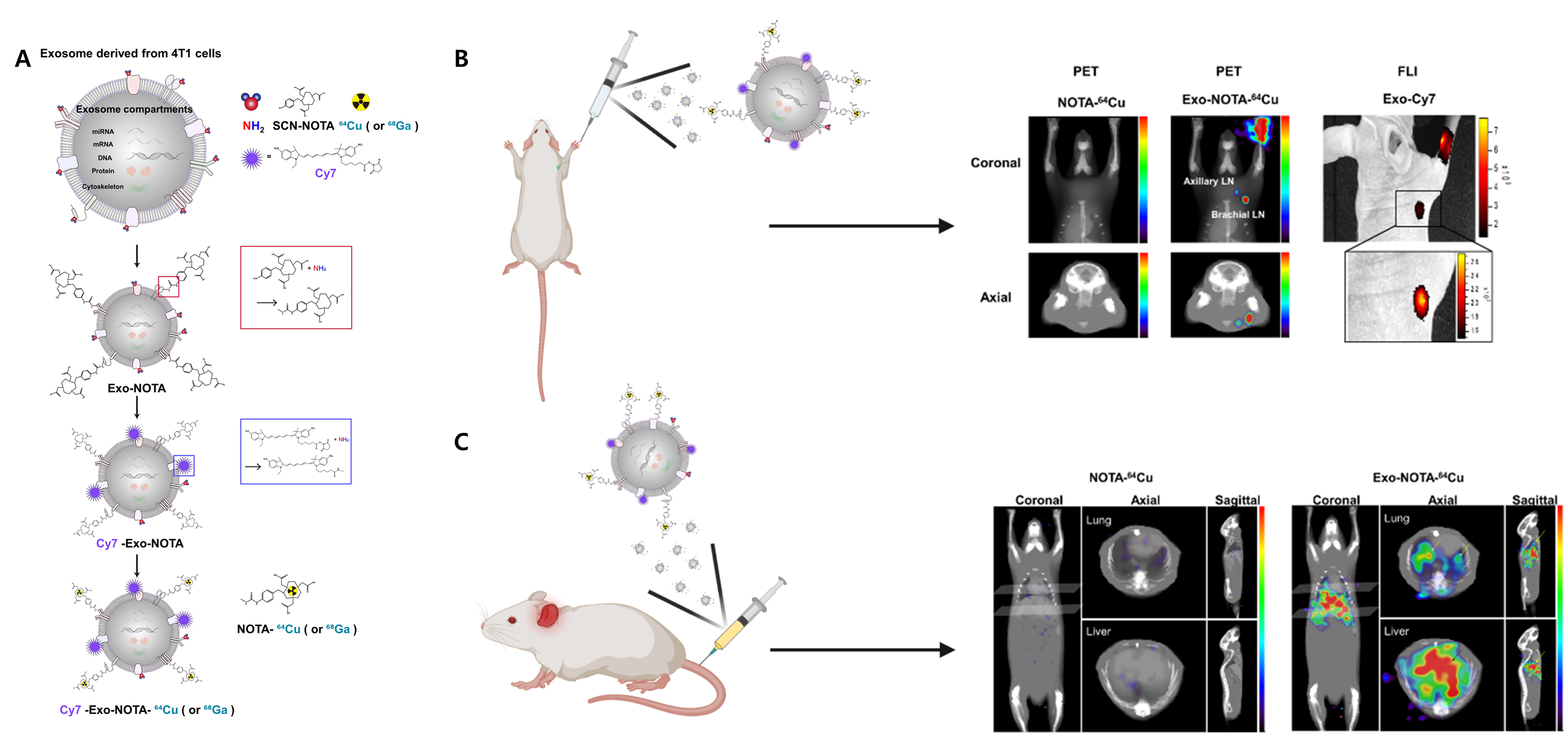
Affiliations
Kyung Oh Jung 1 2 , Young-Hwa Kim 1 3 4 5 , Seock-Jin Chung 1 , Chul-Hee Lee 1 3 , Siyeon Rhee 6 , Guillem Pratx 2 , June-Key Chung 1 , Hyewon Youn 1 3 4 5 7
1 Department of Nuclear Medicine, Seoul National University College of Medicine, Seoul 03080, Korea.
2 Department of Radiation Oncology, Division of Medical Physics, Stanford University School of Medicine, Stanford University, Stanford, CA 94305, USA.
3 Cancer Research Institute, Seoul National University College of Medicine, Seoul 03080, Korea.
4 Medical Research Center, Institute of Radiation Medicine, Seoul National University College of Medicine, Seoul 03080, Korea.
5 Tumor Microenvironment Global Core Research Center, Seoul National University, Seoul 08826, Korea.
6 Department of Biology, Stanford University, Stanford, CA 94305, USA.
7 Cancer Imaging Center, Seoul National University Hospital, Seoul 03080, Korea.
- 키워드
- PET imaging; biodistribution; exosome; optical imaging.
- 연구소개
- 본 연구는 방사성 및 형광이 동시에 표지된 종양세포 유래 엑소좀을 활용하여 림프 및 전신의 영상화에 관한 연구이다. 치료 및 유망한 진단 바이오 마커를 위한 전달 수단으로서 엑소좀의 임상적 사용에 있어서 엑소좀의 생리 학적 분포는 그 유효성과 안전성을 검증하기 위해 필수적이다. 마우스에서 방사성 및 형광 표지된 종양세포 유래 엑소좀은 림프 및 혈액 전이 경로를 통해 주입한 후, 양전자 방출 단층 촬영 (PET) 및 광학 영상장비를 통해 영상화 및 정량화 하였다. 림프 전이 경로에 있는 엑소좀은 감시 림프절에서 특이적으로 관찰되었고 원거리 리프절에서는 관찰되지 않았다, 혈액 전이 경로를 통한 투여 후, 엑소좀은 폐, 간 및 비장에서 명확하게 관찰되었다. 이러한 신속한 표지 방법 및 생리학적 분포에 관한 연구는 향후 엑소좀을 활용한 연구 및 임상 적용에 있어서 유용한 정보를 제공할 수 있을 것으로 기대된다.
- 덧글달기


편집위원
암 엑소좀이 양전자 방출 방사성핵종과 형광 색소를 표지하여 엑소좀의 이동을 동물모델에서 확인한 전임상연구임. 엑소좀 연구의 기반기술로 이용될 수 있어 엑소좀관련 연구자에게 에게 관심을 끌 논문으로 생각됨.
덧글달기닫기2020-12-02 11:15:42
등록