


글로벌 연구동향
분자영상 및 방사화학


![[Theranostics] Cerenkov luminescence imaging of interscapular brown adipose tissue using a TSPO-targeting PET probe in the UCP1 ThermoMouse](/enewspaper/upimages/1665051749admin.JPG) [Theranostics] Cerenkov luminescence imaging of interscapular brown adipose tissue using a TSPO-targeting PET probe in the UCP1 ThermoMouse
[Theranostics] Cerenkov luminescence imaging of interscapular brown adipose tissue using a TSPO-targeting PET probe in the UCP1 ThermoMouse서울의대 / 이석용, 오호림, 윤혜원*
- 출처
- Theranostics
- 등재일
- 2022 Aug 29
- 저널이슈번호
- 12(14):6380-6394
- 내용
Abstract
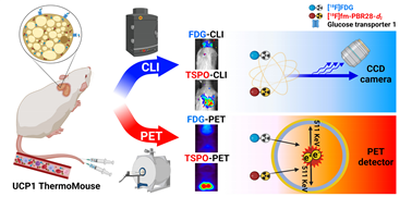
Rationale: [18F]fluorodeoxyglucose-positron emission tomography ([18F]FDG-PET) has been widely used as an imaging technique to measure interscapular brown adipose tissue (iBAT) activity. However, it is challenging to obtain iBAT-specific images using [18F]FDG-PET because increased uptake of [18F]FDG is observed in tumors, muscle, and inflamed tissues. Uncoupling protein 1 (UCP1) in the mitochondrial membrane, a well-known molecular marker of BAT, has been proposed as a useful BAT imaging marker. Recently, the UCP1 ThermoMouse was developed as a reporter mouse for monitoring UCP1 expression and investigating BAT activation. In addition, Translocator protein-18 kDa (TSPO) located in the outer mitochondrial membrane is also overexpressed in BAT, suggesting that TSPO-targeting PET has potential for iBAT imaging. However, there are no studies monitoring BAT using TSPO-targeting PET probes in the UCP1 ThermoMouse. Moreover, the non-invasive Cerenkov luminescence imaging (CLI) using Cerenkov radiation from the PET probe has been proposed as an alternative option for PET as it is less expensive and user-friendly. Therefore, we selected [18F]fm-PBR28-d2 as a TSPO-targeting PET probe for iBAT imaging to evaluate the usefulness of CLI in the UCP1 ThermoMouse.
Methods: UCP1 ThermoMouse was used to monitor UCP1 expression. Western blotting and immunohistochemistry were performed to measure the level of protein expression. [18F]fm-PBR28-d2 and [18F]FDG were used as radioactive probes for iBAT imaging. PET images were acquired with SimPET, and optical images were acquired with IVIS 100.
Results: UCP1 ThermoMouse showed that UCP1 and TSPO expressions were correlated in iBAT. In both PET and CLI, the TSPO-targeting probe [18F]fm-PBR28-d2 was superior to [18F]FDG for acquiring iBAT images. The high molar activity of the probe was essential for CLI and PET imaging. We tested the feasibility of TSPO-targeting probe under cold exposure by imaging with TSPO-PET/CLI. Both signals of iBAT were clearly increased after cold stimulation. Under prolonged isoflurane anesthesia, TSPO-targeting images showed higher signals from iBAT in the short-term than in long-term groups.
Conclusion: We demonstrated that TSPO-PET/CLI reflected UCP1 expression in iBAT imaging better than [18F]FDG-PET/CLI under the various conditions. Considering convenience and cost, TSPO-CLI could be used as an alternative TSPO-PET technique for iBAT imaging.
.
Affiliations
Seok-Yong Lee1,2,3#, Ho Rim Oh1,2,3#, Young-Hwa Kim1,3, Sung-Hwan Bae1,3, Yongseok Lee4, Yun-Sang Lee1,4,5, Byung Chul Lee6,7, Gi Jeong Cheon1,3, Keon Wook Kang1,2,3, Hyewon Youn1,3,4
1. Department of Nuclear Medicine, Seoul National University College of Medicine, Seoul, Republic of Korea
2. Department of Biomedical Sciences, Seoul National University Graduate School, Seoul, Republic of Korea
3. Cancer Research Institute, Seoul National University College of Medicine, Seoul, Republic of Korea
4. Cancer Imaging Center, Seoul National University Hospital, Seoul, Republic of Korea
5. Radiation Medicine Research Institute, Medical Research Center, Seoul National University College of Medicine, Seoul, Republic of Korea
6. Department of Nuclear Medicine, Seoul National University Bundang Hospital, Seongnam, Republic of Korea
7. Center for Nanomolecular Imaging and Innovative Drug Development, Advanced Institutes of Convergence Technology, Seoul National University, Suwon, Republic of Korea
# These authors equally contributed to this research.
- 키워드
- Interscapular brown adipose tissue, UCP1, TSPO, PET, Cerenkov luminescence imaging
- 연구소개
- [18F]FDG-PET은 갈색 지방 조직 (iBAT)의 활성도를 측정하는 일반적인 기법으로 이용되어 오고 있음. 그러나 환자의 종양 및 염증 여부 및 뇌와 심장 등의 섭취가 높아 iBAT의 특이적 영상 획득이 어려움이 있음. 이에 따라 iBAT의 바이오마커로 알려진 UCP1의 발현을 실시간으로 모니터링 할 수 있는 형질전환 마우스 (ThermoMouse) 가 개발 되었음. 또한 TSPO 단백질도 iBAT에 과발현 되어있고 이를 표적하는 PET 프로브를 이용하여 iBAT 영상 획득에 사용되고 있음. Cerenkov luminescence imaging (CLI)는 PET 프로브로부터 방출되는 Cerenkov radation을 optical 장비로 검출하는 영상 기법으로서 경제적이고 이용친화적인 장점으로 PET의 대체 옵션으로 제안되고 있음. 본 연구에서는 UCP1 ThermoMouse에서 기존 [18F]FDG와 TSPO 표적 프로브를 이용해 iBAT 영상 획득의 비교와 PET의 대체 옵션으로서의 CLI의 가능성을 평가하고자 하였음. 본 연구는 기존 [18F]FDG보다 TSPO 표적 프로브가 PET과 CLI 모두 UCP1 발현을 신뢰적으로 반영할 수 있는 iBAT 표적 영상 리간드임을 확인하였고, TSPO-CLI가 TSPO-PET을 효과적으로 대체할 수 있음을 제시하였음.
- 덧글달기


![]()
- COPYRIGHT(C) 2015 한국원자력의학원 전략기획팀 All rights Reserved.
- 문의 : rmwebzine@kirams.re.kr 발행처 : 한국원자력의학원 전략기획팀
- 우) 01812 서울시 노원구 노원로 75 한국원자력의학원 전략기획팀
편집위원
Cerenkov luminescence는 Cerenkov radiation을 이용하여 bioluminescence같은광학영상을 얻는 영상 기술이다. 따라서 F-18 방사성표지자를 이용하면 PET 영상뿐만 아니라 Cerenkov luminescence 영상까지 얻을 수 있다. Brown fat으로 알려진 interscapular brown adipose tissue (iBAT)는 [F-18]FDG PET으로도 일부 영상이 가능하지만, [F-18]FDG는 원래 종양 영상 목적의 방사성의약품이다. 따라서, 최적의 BAT 영상을 위해 이 논문은 Translocator protein-18 kDa (TSPO)를 표적으로 하는 F-18 바이오마커인 [F-18]fm-PBR28-d2를 이용하여 TSPO-PET 및 TSPO-CLI iBAT 영상 결과를 보여주고 있다.
2022-10-06 10:10:28
편집위원
Cerenkov luminescence는 Cerenkov radiation을 이용하여 bioluminescence같은광학영상을 얻는 영상 기술이다. 따라서 F-18 방사성표지자를 이용하면 PET 영상뿐만 아니라 Cerenkov luminescence 영상까지 얻을 수 있다. Brown fat으로 알려진 interscapular brown adipose tissue (iBAT)는 [F-18]FDG PET으로도 일부 영상이 가능하지만, [F-18]FDG는 원래 종양 영상 목적의 방사성의약품이다. 따라서, 최적의 BAT 영상을 위해 이 논문은 Translocator protein-18 kDa (TSPO)를 표적으로 하는 F-18 바이오마커인 [F-18]fm-PBR28-d2를 이용하여 TSPO-PET 및 TSPO-CLI iBAT 영상 결과를 보여주고 있다.
2022-12-06 15:26:15