


글로벌 연구동향
의학물리학


![[IEEE Trans Med Imaging.] DeepRegularizer: Rapid Resolution Enhancement of Tomographic Imaging Using Deep Learning](/enewspaper/upimages/1625212719admin.JPG) [IEEE Trans Med Imaging.] DeepRegularizer: Rapid Resolution Enhancement of Tomographic Imaging Using Deep Learning 딥러닝 기술을 사용하여 단층 이미징의 빠른 해상도 향상 연구
[IEEE Trans Med Imaging.] DeepRegularizer: Rapid Resolution Enhancement of Tomographic Imaging Using Deep Learning 딥러닝 기술을 사용하여 단층 이미징의 빠른 해상도 향상 연구(주)토모큐브, KAIST / 류동훈, 류동민, 민현석*, 박용근*
- 출처
- IEEE Trans Med Imaging.
- 등재일
- 2021 May
- 저널이슈번호
- 40(5):1508-1518. doi: 10.1109/TMI.2021.3058373.
- 내용
-
Abstract
Optical diffraction tomography measures the three-dimensional refractive index map of a specimen and visualizes biochemical phenomena at the nanoscale in a non-destructive manner. One major drawback of optical diffraction tomography is poor axial resolution due to limited access to the three-dimensional optical transfer function. This missing cone problem has been addressed through regularization algorithms that use a priori information, such as non-negativity and sample smoothness. However, the iterative nature of these algorithms and their parameter dependency make real-time visualization impossible. In this article, we propose and experimentally demonstrate a deep neural network, which we term DeepRegularizer, that rapidly improves the resolution of a three-dimensional refractive index map. Trained with pairs of datasets (a raw refractive index tomogram and a resolution-enhanced refractive index tomogram via the iterative total variation algorithm), the three-dimensional U-net-based convolutional neural network learns a transformation between the two tomogram domains. The feasibility and generalizability of our network are demonstrated using bacterial cells and a human leukaemic cell line, and by validating the model across different samples. DeepRegularizer offers more than an order of magnitude faster regularization performance compared to the conventional iterative method. We envision that the proposed data-driven approach can bypass the high time complexity of various image reconstructions in other imaging modalities.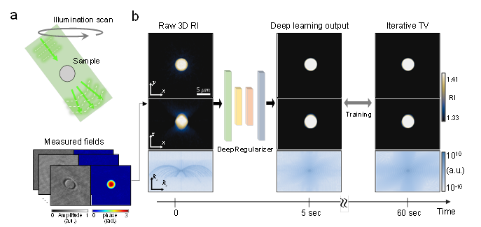
(a) 에서 도식화된 것과 같이 ODT 영상이 제한적인 입사각에서만 촬영되었을 경우, (b) 좌측 (Raw 3D RI) 하단에서 볼 수 있듯이 정보가 없는 주파수 영역을 확인할 수 있다. 제안하는 방식 (Deep learning output)과 기존 복원 알고리즘의 결과 (Iterative TV), 대부분의 정보가 채워진 것을 확인할 수 있는데, 제안하는 방법은 기존 알고리즘에 비해 훨씬 짧은 계산 시간을 보여준다.
DongHun Ryu, Dongmin Ryu, YoonSeok Baek, Hyungjoo Cho, Geon Kim, Young Seo Kim, Yongki Lee, Yoosik Kim, Jong Chul Ye, Hyun-Seok Min, YongKeun Park
- 연구소개
- 토모큐브의 3D 홀로그래피 현미경은 염색과 같은 표지 과정없이 살아있는 세포의 3차원 단층영상을 촬영하여 세포 내 다양항 정량정보를 제공할 수 있습니다. 이는 ODT (Optical diffraction tomography, 광학적 회절 토모그래피)란 기술을 사용하여 측정한 물질 고유의 굴절률값들을 기반으로 제공됩니다. 그러나 ODT 측정에 있어, 여러 제약 상황에 의한 입사광선의 각도가 제약됨에 따라 주파수 공간에서 얻을 수 없는 정보가 존재합니다. 이러한 문제를 해결하기 위하여 굴절률 단층 영상에 대한 물리적 사전정보를 기반으로 계산량이 많은 반복적 복원 알고리즘이 사용되었습니다. 따라서 본 연구에서는 딥러닝 기술을 적용하여 제한된 사전 정보에 특정 상황에서 불안정하지 않고, 고반복의 비효율성을 줄인 효율적인 복원 알고리즘을 제안하였습니다. 본 연구의 결과는 기존 고반복적 알고리즘에 비해 효율적이면서 성능 안정성이 뛰어남을 보여주고 있습니다.
- 덧글달기


![]()
- COPYRIGHT(C) 2015 한국원자력의학원 전략기획팀 All rights Reserved.
- 문의 : rmwebzine@kirams.re.kr 발행처 : 한국원자력의학원 전략기획팀
- 우) 01812 서울시 노원구 노원로 75 한국원자력의학원 전략기획팀