


글로벌 연구동향
핵의학


- [Cells .] Different Expression of Thyroid-Specific Proteins in Thyroid Cancer Cells between 2-Dimensional (2D) and 3-Dimensional (3D) Culture Environment
경북의대 / 오지민, 안병철*
- 출처
- Cells .
- 등재일
- 2022 Nov 10
- 저널이슈번호
- 11(22):3559. doi: 10.3390/cells11223559.
- 내용
Abstract
The two-dimensional (2D) monolayer culture as a conventional method has been widely applied in molecular biology fields, but it has limited capability to recapitulate real cell environments, being prone to misinterpretation with poor prediction of in vivo behavior. Recently, the three-dimensional (3D) spheroid culture has been studied extensively. Spheroids are self-assembled cell aggregates that have biomimicry capabilities. The behavior of thyroid cancer under the 3D spheroid culture environment has been studied; however, there are no reports regarding differences in the degree of thyroid cancer cell differentiation under 2D and 3D culture conditions. This study investigated the expression of thyroid differentiation proteins related to iodide-metabolizing mechanisms in thyroid cancer cells under different culture conditions. Four thyroid cancer cell lines and one thyroid follicular epithelial cell line were grown in adherent 2D cell culture and 3D spheroid culture with agarose-coated plates. We observed changes in proliferation, hypoxia, extracellular matrix (ECM), cytoskeleton, thyroid-specific proteins, and thyroid transcription factors. All cell lines were successfully established in the spheroid following cell aggregation. Proliferation considerably decreased, while hypoxia-inducible factor 1-α(HIF1-α) was promoted in 3D spheroids; moreover, 3D spheroids with thyroid cancers showed diminished thyroid differentiation markers, but thyroid follicular epithelial cells revealed either a maintenance or weak decline of protein expression. We verified that the 3D spheroid culture environment can be similar to in vivo conditions because of its alterations in numerous cellular and functional activities, including morphology, cellular proliferation, viability, hypoxia, ECM, cytoskeleton, and thyroid differentiation, compared to the conventional 2D monolayer culture environment. An in vitro experimental study using 3D spheroid culture is ideal for the faster discovery of new drugs.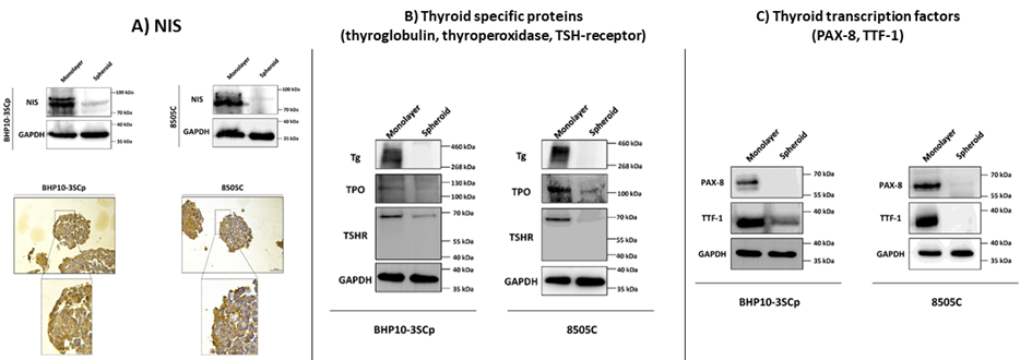
Affiliations
Ji Min Oh 1, Prakash Gangadaran 1 2, Ramya Lakshmi Rajendran 1, Chae Moon Hong 1 3, Jaetae Lee 1 3, Byeong-Cheol Ahn 1 2 3
1Department of Nuclear Medicine, School of Medicine, Kyungpook National University, Daegu 41944, Korea.
2BK21 FOUR KNU Convergence Educational Program of Biomedical Sciences for Creative Future Talents, Department of Biomedical Science, School of Medicine, Kyungpook National University, Daegu 41944, Korea.
3Department of Nuclear Medicine, Kyungpook National University Hospital, Daegu 41944, Korea.
- 키워드
- 3-dimensional culture; Hypoxia; Thyroid differentiation; thyroid cancers.
- 연구소개
- 신약 개발을 위한 시험관 연구는 주로 바닥에 세포를 부착시켜 배양하는 2차원적 기법이 많이 활용되며, 갑상선암의 재분화 약제 선별 연구 또한 통상적으로 2차원적 배양을 이용함. 2차원적 배양기법은 세포가 단일층으로 증식하므로 실제 생체 내에서 세포가 3차원적으로 증식하는 것과는 차이가 나며, 이러한 이질적인 환경은 종양의 미세환경과 유전자 발현에도 영향을 미쳐 동물실험 및 임상 적용에서 다른 결과를 초래할 수 있다는 연구결과가 있음. 본 연구의 궁극적 목표는 다른 배양환경(2차원과 3차원)에서 배양된 갑상선암세포에서 갑상선 분화 단백질 변화차이를 확인하고자 하였음. 2차원 적배양에 비해 3차원적 배양에서는 갑상선암세포의 갑상선 분화 관련 단백인 sodium iodide symporter (NIS), PAX-8, TTF-1, TSH-receptor, thyroperoxidase, thyroglobulin 발현이 감소함. 이는 주로 산소와 영양분 공급이 부족하여 necrotic이 보이는 inner layer에서 단백 발현이 저하됨. 해당 연구는 2차원적 배양과 3차원적 배양에서 갑상선암의 분화 단백 변화를 비교분석함으로써 2차원적 배양에서 주로 행해진 재분화 관련 시험관내 연구에 대해 3차원적 배양법의 활용가능성을 제시함.
- 덧글달기


![]()
- COPYRIGHT(C) 2015 한국원자력의학원 전략기획팀 All rights Reserved.
- 문의 : rmwebzine@kirams.re.kr 발행처 : 한국원자력의학원 전략기획팀
- 우) 01812 서울시 노원구 노원로 75 한국원자력의학원 전략기획팀
편집위원
세포배양을 통한 전임상연구는 2차원에서 배양하는 방식이 이용되는 것이 대부분이다. 전임상연구가 동물실험으로 진행하면 세포가 2차원이 아닌 3차원으로 성장하게 된다. 최근 세포배양 전임상연구에서 3차원 배양을 이용한 연구가 증가되도 있다. 해당 연구는 갑상선암세포주를 2차원 배양과 3차원 배양시 갑상선특이단백의 발현에 차이가 있음을 보여준 전임상 연구이다. 갑상선암의 분화 및 재분화에 대하여 관심있는 기초의학연구자 및 방사성요오드 치료 관심 핵의학의사에게 관심을 끌 흥미로운 연구로 생각된다.
2023-01-06 15:03:41