


글로벌 연구동향
핵의학


![[J Nucl Med.] PET Imaging of System xC - in Immune Cells for Assessment of Disease Activity in Mice and Patients with Inflammatory Bowel Disease](/enewspaper/upimages/1670371219admin.JPG) [J Nucl Med.] PET Imaging of System xC - in Immune Cells for Assessment of Disease Activity in Mice and Patients with Inflammatory Bowel Disease염증성 장질환 진단을 위한 면역 세포의 xC-의 PET 영상 연구
[J Nucl Med.] PET Imaging of System xC - in Immune Cells for Assessment of Disease Activity in Mice and Patients with Inflammatory Bowel Disease염증성 장질환 진단을 위한 면역 세포의 xC-의 PET 영상 연구울산의대 / 서민정, 김예지, 예병덕, 권미나*, 문대혁*
- 출처
- J Nucl Med.
- 등재일
- 2022 Oct
- 저널이슈번호
- 63(10):1586-1591. doi: 10.2967/jnumed.121.263289. Epub 2022 Jan 27.
- 내용
-
Abstract
We aimed to explore whether the imaging of antiporter system xC - of immune cells with (4S)-4-(3-18F-fluoropropyl)-l-glutamate (18F-FSPG) PET can assess inflammatory bowel disease (IBD) activity in murine models and patients (NCT03546868). Methods: 18F-FSPG PET imaging was performed to assess IBD activity in mice with dextran sulfate sodium-induced and adoptive T-cell transfer-induced IBD and a cohort of 20 patients at a tertiary care center in South Korea. Immunohistochemical analysis of system xC - and cell surface markers was also studied. Results: Mice with experimental IBD showed increased intestinal 18F-FSPG uptake and xCT expression in cells positive (+) for CD11c, F4/80, and CD3 in the lamina propria, increases positively associated with clinical and pathologic disease activity. 18F-FSPG PET studies in patients, most of whom were clinically in remission or had mildly active IBD, showed that PET imaging was sufficiently accurate in diagnosing endoscopically active IBD and remission in patients and bowel segments. 18F-FSPG PET correctly identified all 9 patients with superficial or deep ulcers. Quantitative intestinal 18F-FSPG uptake was strongly associated with endoscopic indices of IBD activity. The number of CD68+xCT+ and CD3+xCT+ cells in 22 bowel segments from patients with ulcerative colitis and the number of CD68+xCT+ cells in 7 bowel segments from patients with Crohn disease showed a significant positive association with endoscopic indices of IBD activity. Conclusion: The assessment of system xC - in immune cells may provide diagnostic information on the immune responses responsible for chronic active inflammation in IBD. 18F-FSPG PET imaging of system xC - activity may noninvasively assess the IBD activity.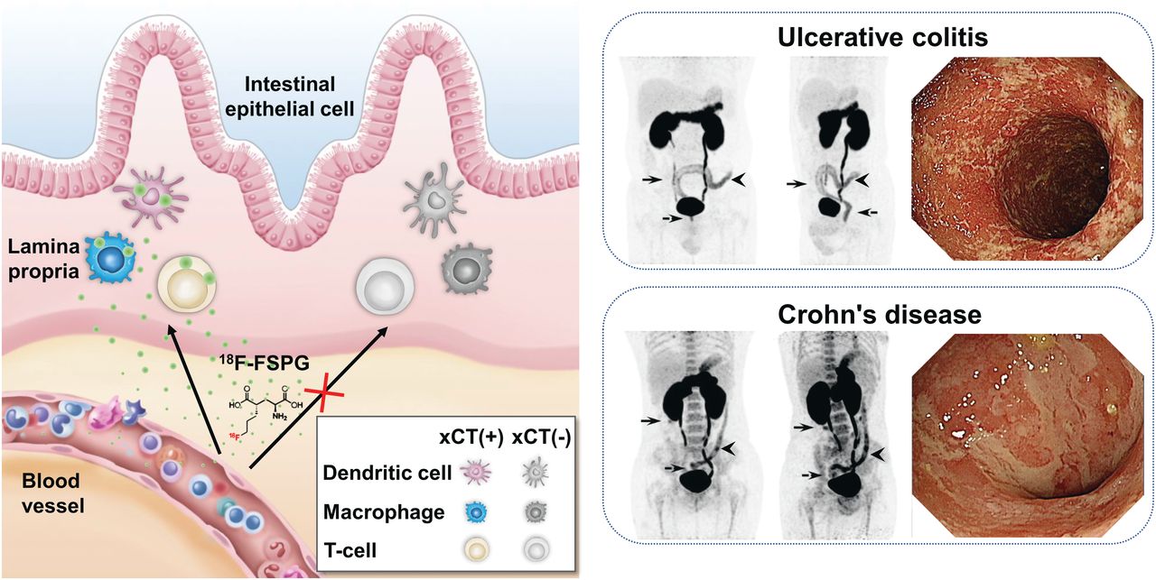
Affiliations
Minjung Seo 1, Yeji Kim 2, Byong Duk Ye 3, Sang Hyoung Park 3, Seog-Young Kim 2, Jin Hwa Jung 2, Sung Wook Hwang 3, Sun Young Chae 4, Dong Yun Lee 4, Sang Ju Lee 4, Seung Jun Oh 4, Jihun Kim 5, Ji Young Kim 6, Sae Jung Na 7, Misung Kim 8, Sang-Yeob Kim 2, Norman Koglin 9, Andrew W Stephens 9, Mi-Na Kweon 10, Dae Hyuk Moon 11
1Department of Nuclear Medicine, Ulsan University Hospital, University of Ulsan College of Medicine, Ulsan, Republic of Korea.
2Department of Convergence Medicine, Asan Medical Center, University of Ulsan College of Medicine, Seoul, Republic of Korea.
3Department of Gastroenterology, Asan Medical Center, University of Ulsan College of Medicine, Seoul, Republic of Korea.
4Department of Nuclear Medicine, Asan Medical Center, University of Ulsan College of Medicine, Seoul, Republic of Korea.
5Department of Pathology, Asan Medical Center, University of Ulsan College of Medicine, Seoul, Republic of Korea.
6Department of Nuclear Medicine, Hanyang University Medical Center, Hanyang University College of Medicine, Seoul, Republic of Korea.
7Department of Radiology, Uijeongbu St. Mary's Hospital, College of Medicine, Catholic University of Korea, Seoul, Republic of Korea.
8Department of Pathology, Ulsan University Hospital, Ulsan, Republic of Korea; and.
9Life Molecular Imaging GmbH, Berlin, Germany.
10Department of Convergence Medicine, Asan Medical Center, University of Ulsan College of Medicine, Seoul, Republic of Korea; dhmoon@amc.seoul.kr mnkweon@amc.seoul.kr.
11Department of Nuclear Medicine, Asan Medical Center, University of Ulsan College of Medicine, Seoul, Republic of Korea; dhmoon@amc.seoul.kr mnkweon@amc.seoul.kr.
- 키워드
- PET; immune cells; inflammatory bowel disease; system xC−.
- 연구소개
- 염증성 장질환(궤양성대장염과 크론병)은 점차 발생이 증가하는 치료가 어려운 만성질환이다. 특히 치료에 있어서 활동성 여부를 평가하는 것이 매우 중요한데, 이를 위해 반드시 대장내시경을 시행하여야 하는 문제점이 있다. 본 연구에서는 염증성 장질환에 대한 전임상 모델 2종 (Dextran sulfate sodium 유도 모델, T세포 이식 모델)과 20명의 환자에서 18F-FSPG PET영상이 장염증의 활동도 평가에서의 유용함과 그 영상 기전이 염증세포의 system xC¯ 활성화에 의한 것이라는 결과를 얻었다. 이러한 결과는 18F-FSPG PET영상법으로 염증성 장질환의 활동도를 비침습적으로 평가할 수 있다는 것을 의미한다.
- 덧글달기


![]()
- COPYRIGHT(C) 2015 한국원자력의학원 전략기획팀 All rights Reserved.
- 문의 : rmwebzine@kirams.re.kr 발행처 : 한국원자력의학원 전략기획팀
- 우) 01812 서울시 노원구 노원로 75 한국원자력의학원 전략기획팀
편집위원
새로운 PET용 방사성의약품인 (4S)-4-(3-18F-fluoropropyl)-l-glutamate (18F-FSPG) 을 이용하여 전임상 임상연구를 통해 염증성 잘질환 평가에 대한 연구 결과를 보여준 논문이다. 면역세포의 System XC−를 18F-FSPG PET 영상화 하여 염증성 잘 질환의 평가에 이용가능함을 보여준 연구임. 대장질환 전문가와 핵의학 임상가 뿐만 아니라 방사화학자에게 관심을 끌 흥미로운 연구로 생각된다.
2022-12-06 15:12:06