


글로벌 연구동향
핵의학


- [Sci Rep.] Radioiodine labeling and in vivo trafficking of extracellular vesicles
경북의대 / 홍채문, 안병철*
- 출처
- Sci Rep.
- 등재일
- 2021 Mar 3
- 저널이슈번호
- 11(1):5041. doi: 10.1038/s41598-021-84636-5.
- 내용
Abstract
Biodistribution and role of extracellular vesicles (EVs) are still largely unknown. Reliable tracking methods for EVs are needed. In this study, nuclear imaging using radioiodine were developed and applied for tracking EVs derived from cell lines. EVs were obtained from supernatant of thyroid cancer cell (Cal62) and natural killer cells (NK92-MI) using sequential ultracentrifuges. Sulfosuccinimidyl-3-(4-hydroxypheynyl) propionate were labeled to membrane of Cal62 and NK92-MI cell derived EVs, then the EVs were labeled with radioiodine (I-131 and I-125) using pre-coated iodination tubes (RI-EVs). In vivo gamma camera images were obtained after intravenous injection of the RI-EVs, and ex vivo biodistribution study was also performed. EVs were labeled with radioiodine and radiochemical purity of the RI-EV was more than 98%. Results of nanoparticle tracking analysis and electron microscopy showed that there was no significant difference in EVs before and after the radioiodine labeling. After intravenous injection of RI-EVs to mice, gamma camera imaging well visualized the real-time biodistribution of the RI-EVs. RI-EVs were mainly visualized at liver, spleen, and lung. Nuclear imaging system of EVs derived from thyroid cancer and NK cells using radioiodine labeling of the EVs was established. Thus, this system might be helpful for in vivo tracking of EVs.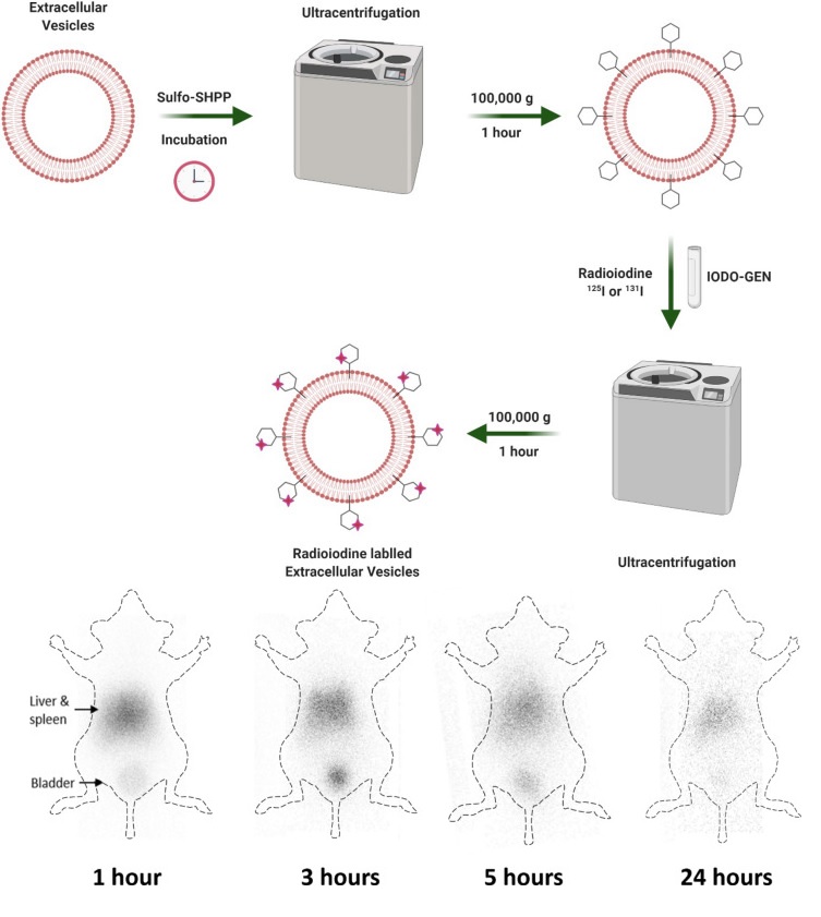
Affiliations
Chae Moon Hong 1 2 , Prakash Gangadaran 1 3 , Ji Min Oh 1 , Ramya Lakshmi Rajendran 1 , Arunnehru Gopal 1 , Liya Zhu 1 , Byeong-Cheol Ahn 4 5
1 Department of Nuclear Medicine, School of Medicine, Kyungpook National University, 130 Dongdeok-ro, Jung Gu, Daegu, 41944, Republic of Korea.
2 Department of Nuclear Medicine, Kyungpook National University Hospital, Daegu, Republic of Korea.
3 BK21 FOUR KNU Convergence Educational Program of Biomedical Sciences for Creative Future Talents, School of Medicine, Kyungpook National University, Daegu, Republic of Korea.
4 Department of Nuclear Medicine, School of Medicine, Kyungpook National University, 130 Dongdeok-ro, Jung Gu, Daegu, 41944, Republic of Korea. abc2000@knu.ac.kr.
5 Department of Nuclear Medicine, Kyungpook National University Hospital, Daegu, Republic of Korea. abc2000@knu.ac.kr.
- 연구소개
- 최근 활발한 연구가 진행되고 있는 세포 외 소포 (Extracellular vesicle)에 방사성동위원소인 I-131 또는 I-125를 세포 외 소포 표면 two-step 방법으로 표지하여 세포외 소포의 생체내 분포를 비침습적으로 영상화 가능성을 보여준 연구입니다. 감마카메라 영상 분석이 biodistribution 결과를 잘 반영하였습니다.
- 덧글달기


![]()
- COPYRIGHT(C) 2015 한국원자력의학원 전략기획팀 All rights Reserved.
- 문의 : rmwebzine@kirams.re.kr 발행처 : 한국원자력의학원 전략기획팀
- 우) 01812 서울시 노원구 노원로 75 한국원자력의학원 전략기획팀
편집위원
세포외 소포의 전임상 및 임상연구에는 세포외 소포의 비침습적 영상기술의 개발이 필수적이다. 세포외 소포에 방사성요오드를 표지하는 방법에 대한 연구로 세포외 소포 관련 연구자 및 세포 및 세포 유래 소표에 방사성핵종을 표지하고자 하는 연구자에게 관심을 끌 연구로 생각됨.
2021-05-06 15:31:42
편집위원2
엑소좀을 핵의학에서 흔히쓰는 Iodine으로 label하여 in vivo distribution을 보는 흥미로운 기초연구라 생각합니다.
2021-05-06 15:34:58