
글로벌 연구동향
의학물리학


![[Phys Med Biol.] Development of a deep neural network for generating synthetic dual-energy chest x-ray images with single x-ray exposure.](/enewspaper/upimages/1564468042admin.JPG) 2019년 08월호
2019년 08월호
[Phys Med Biol.] Development of a deep neural network for generating synthetic dual-energy chest x-ray images with single x-ray exposure.단일 X-선을 이용하여 이중에너지 흉부 X-선 합성 영상 생성을 위한 심 신경 네트워크 개발 연구연세대 / 이동훈, 김휘영, 최병욱*, 김희중*
- 출처
- Phys Med Biol.
- 등재일
- 2019 May 31
- 저널이슈번호
- 64(11):115017. doi: 10.1088/1361-6560/ab1cee.
- 내용
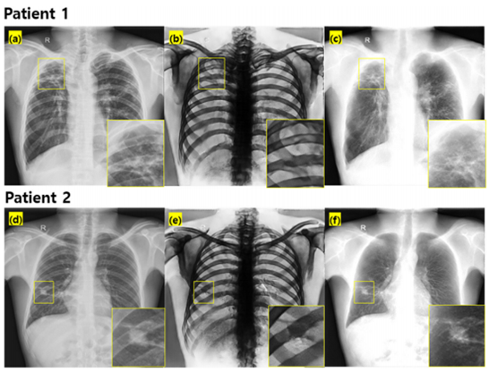
위의 그림은 중국 선전 (Shenzhen)에서 촬영된 흉부 X-선 영상에 제안한 기술을 적용한 결과입니다. 이중에너지를 사용하지 않았음에도, 제안한 기술을 사용함으로써 연부 조직과 뼈 성분을 구분하여 영상화할 수가 있었습니다. 첫 번째 환자는 Right Lung Apex에 Tuberculosis 가 있는 환자였고, 두 번째 환자는 Right Lung에 Nodule이 있는 환자였습니다. 제안한 기술은 병변의 신호에는 왜곡을 주지 않았고, 효과적으로 뼈 성분을 제거하여 병변 진단에 도움을 줄 수 있을 것으로 기대됩니다.
Abstract
Dual-energy chest radiography (DECR) is a medical imaging technology that can improve diagnostic accuracy. This technique can decompose single-energy chest radiography (SECR) images into separate bone- and soft tissue-only images. This can, however, double the radiation exposure to the patient. To address this limitation, we developed an algorithm for the synthesis of DECR from a SECR through deep learning. To predict high resolution images, we developed a novel deep learning architecture by modifying a conventional U-net to take advantage of the high frequency-dominant information that propagates from the encoding part to the decoding part. In addition, we used the anticorrelated relationship (ACR) of DECR for improving the quality of the predicted images. For training data, 300 pairs of SECR and their corresponding DECR images were used. To test the trained model, 50 DECR images from Yonsei University Severance Hospital and 662 publicly accessible SECRs were used. To evaluate the performance of the proposed method, we compared DECR and predicted images using a structural similarity approach (SSIM). In addition, we quantitatively evaluated image quality calculating the modulation transfer function and coefficient of variation. The proposed model selectively predicted the bone- and soft tissue-only CR images from an SECR image. The strategy for improving the spatial resolution by ACR was effective. Quantitative evaluation showed that the proposed method with ACR showed relatively high SSIM (over 0.85). In addition, predicted images with the proposed ACR model achieved better image quality measures than those of U-net. In conclusion, the proposed method can obtain high-quality bone- and soft tissue-only CR images without the need for additional hardware for double x-ray exposures in clinical practice.
Author informationLee D1, Kim H, Choi B, Kim HJ.
1
Department of Radiation Convergence Engineering, Research Institute of Health Science, Yonsei University, 1 Yonseidae-gil, Wonju, Gangwon, Republic of Korea.
- 연구소개
- 흉부 진단에 도움이 되는 이중에너지 X-ray 영상의 구현은 환자한테 두 번의 X-ray 조사가 필요하다는 문제점이 있었습니다. 이 때문에, 빠른 시간에 두 번의 X-ray 조사를 위한 Hardware 기술이 필요했고, 환자한테 전달되는 선량이 증가하고 Motion artifact가 발생되는 부작용이 문제점이었습니다. 본 연구는 기존의 이중에너지의 문제점이었던 두 두 번의 X-ray 조사 없이 일반 흉부 X-ray 영상에서 딥러닝을 통해 이중에너지 흉부 X-ray 영상의 구현에 관한 내용입니다.
- 덧글달기


편집위원
AI는 방사선의할 뿐만 아니라 응용되지 않는 분야가 거의 없을 만큼 다양하게 응용되고 있다. 방사선의학 안에서도 매우 다양한 분야에서 적용되고 있는데, 본 논문은 영상의학 분야에서 기존 CR 한 장으로 dual energy CR을 얻는 효과를 낼 수 있는 방법을 보였다는 점에서 흥미로왔다. 정확도를 높이고 유용성이 확인되어 실제 임상에 적용되기까지는 앞으로도 시간이 더 필요하겠지만 좋은 시도와 좋은 결과로 생각된다.
덧글달기닫기2019-07-18 15:27:04
등록