
글로벌 연구동향
핵의학


![[Nucleic Acids Res.] Discovery of molecular features underlying the morphological landscape by integrating spatial transcriptomic data with deep features of tissue images](/enewspaper/upimages/1627982148admin.JPG) 2021년 08월호
2021년 08월호
[Nucleic Acids Res.] Discovery of molecular features underlying the morphological landscape by integrating spatial transcriptomic data with deep features of tissue images 공간적 유전자특성에 따른 조직의 형태발현 분석서울의대 / 배성우, 최홍윤*, 이동수*
- 출처
- Nucleic Acids Res.
- 등재일
- 2021 Jun 4
- 저널이슈번호
- 49(10):e55. doi: 10.1093/nar/gkab095.
- 내용
-
Abstract
Profiling molecular features associated with the morphological landscape of tissue is crucial for investigating the structural and spatial patterns that underlie the biological function of tissues. In this study, we present a new method, spatial gene expression patterns by deep learning of tissue images (SPADE), to identify important genes associated with morphological contexts by combining spatial transcriptomic data with coregistered images. SPADE incorporates deep learning-derived image patterns with spatially resolved gene expression data to extract morphological context markers. Morphological features that correspond to spatial maps of the transcriptome were extracted by image patches surrounding each spot and were subsequently represented by image latent features. The molecular profiles correlated with the image latent features were identified. The extracted genes could be further analyzed to discover functional terms and exploited to extract clusters maintaining morphological contexts. We apply our approach to spatial transcriptomic data from different tissues, platforms and types of images to demonstrate an unbiased method that is capable of obtaining image-integrated gene expression trends.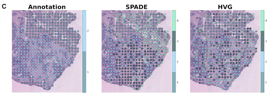
그림 : 전립선암 조직에 SPADE 방법을 적용하여 형태적 특성을 반영하는 유전자를 추출하고 조직의 각 영역을 군집화 하였습니다. SPADE는 기존의 highly variable gene(HVG)를 이용한 방법과 비교하였을 때 종양과 비종양 조직을 높은 정확도로 (SPADE: 90.51%, HVG: 60.40%) 구분할 수 있었습니다. 상기 조직은 Berglund E. et al. Nat. Commun. 2018; 9:2419. 논문에서 유래한 데이터이며, 조직 각 영역이 종양인지 여부를 판단한 병리학적 annotation 정보를 활용하여 진단 정확도를 산출하였습니다.

그림 : 면역 형광 염색을 시행한 mouse brain 조직에 SPADE를 적용하여 조직의 각 영역을 군집화 하였습니다. SPADE는 기존의 HVG 방법과 비교하였을 때 피질층과 피질하 구조물을 효과적으로 구획할 수 있었습니다.
Affiliations
Sungwoo Bae 1 2 , Hongyoon Choi 2 3 , Dong Soo Lee 1 2 3
1 Department of Molecular Medicine and Biopharmaceutical Sciences, Graduate School of Convergence Science and Technology, Seoul National University, Seoul, Republic of Korea.
2 Department of Nuclear Medicine, Seoul National University Hospital, Seoul, Republic of Korea.
3 Department of Nuclear Medicine, Seoul National University College of Medicine, Seoul, Republic of Korea.
- 연구소개
- 차세대 염기서열 분석과 조직에서의 위치 바코딩 기술을 결합한 공간 전사체학 방법이 상용화되면서 공간적인 유전자 발현과 그 상호작용을 분석할 수 있는 플랫폼이 마련되었습니다. 공간 전사체학 데이터를 활용하여 공간적으로 발현이 상이한 유전자를 탐색하는 여러 분석 도구들이 개발되었지만, 발현 정보와 함께 얻어지는 조직의 이미지 정보를 통합하는 멀티 오믹스 분석 기법은 확립되지 않은 상태입니다. 조직 각 위치에서의 형태적인 특성을 정량적으로 산출하여 유전자 발현과의 연관성을 탐색한다면 다양한 질병 모델에서 조직의 구조와 연결된 병태생리를 찾아나가는 데에 큰 도움이 될 것입니다. 이에 저희 연구팀에서는 공간 전사체학 분석에서 얻어진 조직 각 위치에서의 유전자 발현 정보와 이미지 특성을 통합하는 분석 방법(SPADE)을 제안하였습니다. ImageNet Challenge에서 사용되었던 컨볼루션 신경망인 VGG-16을 이용하여 조직의 이미지 특성을 추출하고 주성분 분석법(PCA)으로 차원을 축소하여 조직의 형태학적 특성과 연관된 유전자를 선별해내게 됩니다. 그에 더하여 조직의 이미지 특성을 반영하는 유전자 묶음이 어떤 기능과 관련되어 있는지 기존의 gene ontology 용어로 표현하였으며 묶음 유전자의 발현 관점에서 조직의 각 영역을 군집화 하였습니다. 상기 SPADE 방법을 공공데이터에 적용하여 검증을 시행하였습니다. 첫 번째로, H&E 염색한 사람의 유방암 및 전립선암 조직에서 종양과 기질에 차등적으로 발현된 유전자와 그 기능을 탐색할 수 있었습니다. 또한, 전립선 조직에서 암과 그 이외의 조직을 높은 정확도로 구획하였습니다. 뿐만 아니라, 면역 형광 염색을 시행한 mouse의 뇌에도 적용하여 여러 피질층과 피질하 구조물을 효과적으로 구분해낼 수 있었습니다. 이로써, SPADE는 여러 공간 전사체학 데이터에서 조직의 형태적인 특성을 반영하는 유전자를 추출하고 기능적인 의미를 탐색하는 유용한 도구로 사용될 수 있겠습니다.
- 덧글달기
- 이전글 [Medicine (Baltimore)] Methodological considerations of evaluating the rate of presynaptic dopaminergic denervation in Parkinson disease with radiotracers: Analysis of the PPMI data
- 다음글 [J Clin Endocrinol Metab.] Radioactive Iodine Treatment for Children and Young Adults with Thyroid Cancer in South Korea: A Population-based Study


