
글로벌 연구동향
핵의학


![[Sci Rep.] Usefulness of 68 Ga-DOTATOC PET/CT to localize the culprit tumor inducing osteomalacia](/enewspaper/upimages/1615177475admin.JPG) 2021년 03월호
2021년 03월호
[Sci Rep.] Usefulness of 68 Ga-DOTATOC PET/CT to localize the culprit tumor inducing osteomalacia울산의대 / 이동윤, 류진숙*
- 출처
- Sci Rep.
- 등재일
- 2021 Jan 19
- 저널이슈번호
- 11(1):1819. doi: 10.1038/s41598-021-81491-2.
- 내용
Abstract
Tumor-induced osteomalacia (TIO) is an uncommon paraneoplastic syndrome presenting with sustained hypophosphatemia. Treatment of choice is removal of the tumor causing the TIO, but identification of the culprit tumor by routine imaging is challenging. This study aimed to assess the usefulness of somatostatin receptor imaging, called 68Ga-DOTATOC PET/CT, in the management of patients with TIO. Twelve patients who were suspected of having TIO underwent 68Ga-DOTATOC PET/CT. Lesion detectability and maximum standardized uptake value (SUVmax) were determined and retrospectively compared with the clinical/imaging surveillance and histopathologic diagnosis. The median duration of suspected TIO with hypophosphatemia was 7.8 years (range 2.1-21.0). Conventional radiologic and/or nuclear medicine images failed to identify the culprit tumors. However, 68Ga-DOTATOC PET/CT scans showed that 8 of the 12 patients had positive lesions, suggesting the presence of focal culprit tumors. The SUVmax of positive tumors was 1.9-45.7 (median: 11.5). Six skeletal lesions and two extra-skeletal lesions were identified. Seven of the lesions were pathologically confirmed as potential culprits of TIO. Hypophosphatemia was resolved in five patients who underwent lesion excision. The 68Ga-DOTATOC PET/CT is a useful whole-body imaging modality for the detection of causative tumors in patients with suspected TIO.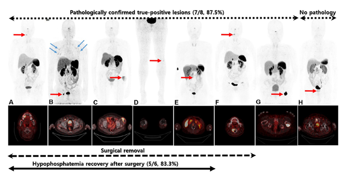
Affiliations
Dong Yun Lee 1 , Seung Hun Lee 2 , Beom-Jun Kim 2 , Wanlim Kim 3 , Pil Whan Yoon 3 , Sang Ju Lee 1 , Seung Jun Oh 1 , Jung-Min Koh 2 , Jin-Sook Ryu 4
1 Department of Nuclear Medicine, Asan Medical Center, University of Ulsan College of Medicine, Seoul, South Korea.
2 Division of Endocrinology and Metabolism, Asan Medical Center, University of Ulsan College of Medicine, Seoul, South Korea.
3 Department of Orthopaedic Surgery, Asan Medical Center, University of Ulsan College of Medicine, Seoul, South Korea.
4 Department of Nuclear Medicine, Asan Medical Center, University of Ulsan College of Medicine, Seoul, South Korea. jsryu2@amc.seoul.kr.
- 연구소개
- 종양성 골연화증이란 간엽성 종양 (mesenchymal tumor) 에서 과다 분비되는 FGF-23이라는 호르몬에 의해 저인산혈증과 골연화증이 특징적으로 유발되는 매우 드문 부종양성 증후군입니다. 이 질환의 근본적인 치료는 뼈나 연조직에 주로 위치하는 간엽성 종양을 제거하는 것이나, MRI와 18F-FDG PET/CT를 포함한 기존의 해부학적/기능적 영상으로는 종양을 정확하게 찾아내는데 많은 어려움이 있었습니다. 그러나 소마토스타틴 수용체가 이 종양에서 과다 발현한다는 데에 착안하여 68Ga-DOTA 기반의 소마토스타틴 유사체 PET/CT 영상을 통해 종양을 효율적이고 정확하게 진단할 수 있다는 발표가 최근에 보고되었습니다. 이에 본원에서도 68Ga‑DOTATOC PET/CT의 임상 도입 후 짧게는 2년, 길게는 21년 동안 이 질환으로 고생하던 환자들의 병변을 정확하게 찾아내고 제거하여 저인산혈증을 포함한 부종양성 증후군을 완전히 관해 시킬 수 있었음을 이번 논문에 보고하였습니다.
- 덧글달기


