


글로벌 연구동향
핵의학


![[Eur Radiol.] PET/CT features discriminate risk of metastasis among single-bone FDG lesions detected in newly diagnosed non-small-cell lung cancer patients.](/enewspaper/upimages/1559110257admin.JPG) [Eur Radiol.] PET/CT features discriminate risk of metastasis among single-bone FDG lesions detected in newly diagnosed non-small-cell lung cancer patients.
[Eur Radiol.] PET/CT features discriminate risk of metastasis among single-bone FDG lesions detected in newly diagnosed non-small-cell lung cancer patients.성균관의대 / 임채홍, 이경한*
- 출처
- Eur Radiol.
- 등재일
- 2019 Apr
- 저널이슈번호
- 29(4):1903-1911. doi: 10.1007/s00330-018-5764-9. Epub 2018 Oct 12.
- 내용
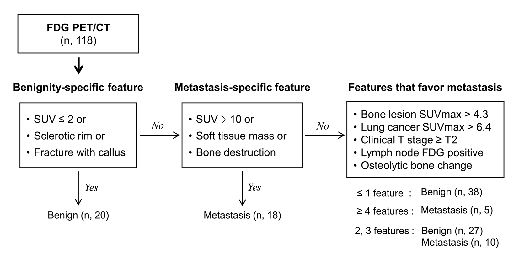
그림) Flowchart for differential diagnosis of single-bone FDG lesions in 118 newly diagnosed NSCLC patients. Using this method, PET/CT features correctly recognized 25 metastatic and 56 benign bone lesions. Only 37 bone lesions remained that required further evaluation for differentiation
Abstract
OBJECTIVES:
We investigated the capacity of fluorodeoxyglucose (FDG) PET/CT features for stratifying probability of metastasis for single-bone FDG lesions in non-small-cell lung cancer (NSCLC).METHODS:
Subjects were 118 newly diagnosed NSCLC patients with a solitary bone FDG lesion and no evidence of other distant metastasis based on PET/CT, brain MRI, and contrast-enhanced chest CT. Bone lesion SUVmax and CT findings, primary tumor SUVmax, clinical T stage, and N stage were analyzed.RESULTS:
The bone lesions were determined by biopsy, characteristic MRI findings and clinical follow-up to be metastatic in 33 (28.0%) and benign in 85 cases (72.0%). A cutoff bone SUVmax of 4.3 showed good diagnostic performance (81.8% sensitivity, 84.7% specificity, and 83.9% accuracy), but there was considerable overlap. Bone lesion PET/CT features of SUVmax ≤ 2, osteosclerotic rim or fracture correctly diagnosed 20/20 benign, while SUVmax > 10, soft-tissue mass or bone destruction correctly diagnosed 18/18 metastatic cases. In the remaining 80 cases, bone features of SUVmax > 4.3 and osteolytic change, and lung tumor features of SUVmax > 6.4, ≥ T2 stage (n = 70), and ≥ N1 stage (n = 43) favored metastasis. The presence of one or less of these features correctly diagnosed 38/38 benign, while the presence of four or more features correctly diagnosed 5/5 metastatic cases. The 37 cases with two or three features had either benign (n = 27) or metastatic bone disease (n = 10).CONCLUSION:
Combining bone lesion and lung tumor PET/CT features can help stratify risk of bone metastasis in these patients.KEY POINTS:
• In NSCLC with a single-bone FDG lesion, lesion SUVmaxis useful for differential diagnosis. • CT features of the single-bone FDG lesions provide additional diagnostic value. • High NSCLC SUVmax, greater T stage, and FDG positive nodes also favor metastasis.
Author informationLim CH1, Ahn TR2, Moon SH1, Cho YS1, Choi JY1, Kim BT1, Lee KH3.
1
Department of Nuclear Medicine, Samsung Medical Center, Sungkyunkwan University School of Medicine, 81 Irwon-ro, Gangnam-gu, Seoul, 06351, South Korea.
2
Department of Radiology, Samsung Medical Center, Sungkyunkwan University School of Medicine, Seoul, 06351, South Korea.
3
Department of Nuclear Medicine, Samsung Medical Center, Sungkyunkwan University School of Medicine, 81 Irwon-ro, Gangnam-gu, Seoul, 06351, South Korea. khnm.lee@samsung.com.
- 키워드
- Bone metastasis; Fluorodeoxyglucose; Lung cancer; Positron-emission tomography
- 연구소개
- 비소세포성 폐암으로 진단 받은 환자의 병기 설정을 위해 촬영한 FDG PET/CT영상에서 원격전이로 의심되는 섭취가 뼈에 한 개 보였을 때 이것의 판단은 어렵지만 환자의 치료와 병기를 바꾸기 때문에 굉장히 중요합니다. 본 연구는 이러한 섭취가 보였을 때, 실제 뼈전이의 가능성이 어느 정도나 될지 FDG PET/CT의 정보를 통하여 예측해보고자 한 연구 논문입니다. 이번 연구는 핵의학과 의사들의 판독에 유용한 정보를 줄 뿐 아니라 더 나아가 임상의들이 비소세포성 폐암환자의 치료방향을 결정하는데 있어서 도움을 줄 것이라 생각합니다.
- 덧글달기


![]()
- COPYRIGHT(C) 2015 한국원자력의학원 전략기획팀 All rights Reserved.
- 문의 : rmwebzine@kirams.re.kr 발행처 : 한국원자력의학원 전략기획팀
- 우) 01812 서울시 노원구 노원로 75 한국원자력의학원 전략기획팀