


글로벌 연구동향
핵의학


![[Eur J Nucl Med Mol Imaging.] Dual-time point 18F-FDG PET/CT for the staging of oesophageal cancer: the best diagnostic performance by retention index for N-staging in non-calcified lymph nodes.](/enewspaper/upimages/1535432927admin.JPG) [Eur J Nucl Med Mol Imaging.] Dual-time point 18F-FDG PET/CT for the staging of oesophageal cancer: the best diagnostic performance by retention index for N-staging in non-calcified lymph nodes.
[Eur J Nucl Med Mol Imaging.] Dual-time point 18F-FDG PET/CT for the staging of oesophageal cancer: the best diagnostic performance by retention index for N-staging in non-calcified lymph nodes.서울의대 / 박소현, 강창현*, 천기정*
- 출처
- Eur J Nucl Med Mol Imaging.
- 등재일
- 2018 Jul
- 저널이슈번호
- 45(8):1317-1328. doi: 10.1007/s00259-018-3981-8. Epub 2018 Mar 3.
- 내용
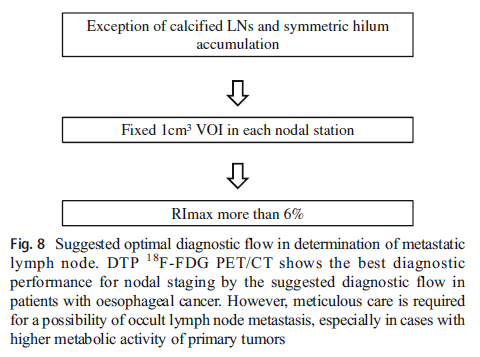
이중촬영 F-18 FDG PET/CT를 이용한 최적의 진단 흐름도. 석회화를 보이고, lung hilar 부위의 대칭적인 F-18 FDG 섭취를 보이는 림프절을 제외하고, 1cm3 의 VOI를 이용하여 SUVmax를 초기/지연촬영에 있어서 각각 측정 한 뒤 RImax가 6%이상인 경우를 전이성 림프절이라고 생각하였을 때, 전이성 림프절 예측에 최적의 결과를 보였다.
Abstract
PURPOSE:
The purpose of this study is to investigate the role of dual time point (DTP) 18F-FDG PET/CT in the staging of oesophageal cancer, especially in lymph node metastasis.METHODS:
A total of 35 patients with oesophageal squamous cell carcinoma who underwent surgical treatment without neoadjuvant chemotherapy were enrolled as a test set and another 19 patients were enrolled as a validation set. The DTP PET/CT scans were obtained in dual time points at 60 and 120 min each, following the administration of 18F-FDG. Visual analysis was performed and semiquantitative analysis was performed using several PET parameters such as maximal standardized uptake values (SUVmax), peak SUV (SUVpeak) and retention indexes using SUVmax (RImax) and SUVpeak (RIpeak).RESULTS:
Primary oesophageal lesions exhibited a significant difference for SUVmax at each time point scan (ANOVA, p < 0.001). For nodal staging, a total of 276 non-calcified nodal stations of the test set were evaluated. Sensitivity, specificity and accuracy of visual analysis were 32.0% (8 of 25), 96.8% (243 of 251) and 90.9% (251 of 276) in the test set. Using ROC analysis, RImax had the largest area under the curve (AUC) to detect metastatic lymphadenopathy at the optimal cut-off value of 6% (AUC 0.853, P < 0.001) in the test set (sensitivity, specificity and accuracy; 80.0% (20 of 25), 94.8% (238 of 251) and 93.5% (258 of 276)). In the validation set (179 non-calcified nodal stations), sensitivity, specificity and accuracy of RImax at the optimal cut-off of 6% were 71.4% (5 of 7), 99.4% (171 of 172) and 98.4% (176 of 179), whereas those of visual analysis were 14.3% (1 of 7), 98.8% (170 of 172) and 95.5% (171 of 179).CONCLUSIONS:
The best diagnostic performance of nodal staging in patients with oesophageal cancer was achieved by application of RImax with a cut-off of more than 6% on DTP 18F-FDG PET/CT with the exclusion of calcified lymph nodes. Optimal clinical management in surgically-candidate oesophageal cancer patients could be achieved using the diagnostic flow on DTP 18F-FDG PET/CT.
Author informationPark S1,2, Paeng JC2,3, Kang CH4,5, Cheon GJ6,7,8, Kang KW2,3,9, Chung JK2,3,9, Lee DS2,10.
1
Department of Nuclear Medicine, National Cancer Center, Seoul, South Korea.
2
Department of Nuclear Medicine, Seoul National University Hospital, College of Medicine, 101 Daehangro, Jongro-gu, Seoul, 110-744, South Korea.
3
Radiation Medicine Research Institute, Seoul National University College of Medicine, Seoul, South Korea.
4
Department of Thoracic and Cardiovascular Surgery, Seoul National University Hospital, College of Medicine, 101 Daehangro, Jongro-gu, Seoul, 110-744, South Korea. chkang@snu.ac.kr.
5
Cancer Research Institute, Seoul National University, Seoul, South Korea. chkang@snu.ac.kr.
6
Department of Nuclear Medicine, Seoul National University Hospital, College of Medicine, 101 Daehangro, Jongro-gu, Seoul, 110-744, South Korea. larrycheon@gmail.com.
7
Radiation Medicine Research Institute, Seoul National University College of Medicine, Seoul, South Korea. larrycheon@gmail.com.
8
Cancer Research Institute, Seoul National University, Seoul, South Korea. larrycheon@gmail.com.
9
Cancer Research Institute, Seoul National University, Seoul, South Korea.
10
Department of Molecular Medicine and Biopharmaceutical Sciences, Graduate School of Convergence Science and Technology, Seoul National University, Seoul, South Korea.http://www.rmwebzine.re.kr/newshome/mtnmain.php?mtnkey=articleview&mkey=scatelist&mkey2=76&aid=2755
- 키워드
- 18F-FDG PET/CT; Dual-time point PET/CT; Oesophageal cancer; Staging
- 연구소개
- 본 논문은 식도암 병기 설정에 있어서 이중 촬영 F-18 FDG PET/CT의 유용성에 관한 논문으로서, 초기 촬영과 지연 촬영의 섭취를 비교한 retention index 와 석회화되지 않은 림프절을 기준으로 하였을 때 우수한 진단성능을 보였다.
- 덧글달기
- 이전글 [Eur J Nucl Med Mol Imaging.] Preoperative [18F]FDG PET/CT tumour heterogeneity index in patients with uterine leiomyosarcoma: a multicentre retrospective study.
- 다음글 [Eur J Nucl Med Mol Imaging.] The role of 18F-FP-CIT PET in differentiation of progressive supranuclear palsy and frontotemporal dementia in the early stage.


![]()
- COPYRIGHT(C) 2015 한국원자력의학원 전략기획팀 All rights Reserved.
- 문의 : rmwebzine@kirams.re.kr 발행처 : 한국원자력의학원 전략기획팀
- 우) 01812 서울시 노원구 노원로 75 한국원자력의학원 전략기획팀