
글로벌 연구동향
방사선종양학


![[BMC Cancer.] Simple calculation using anatomical features on pre-treatment verification CT for bladder volume estimation during radiation therapy for rectal cancer](/enewspaper/upimages/1606889067admin.JPG) 2020년 12월호
2020년 12월호
[BMC Cancer.] Simple calculation using anatomical features on pre-treatment verification CT for bladder volume estimation during radiation therapy for rectal cancer연세의대, 한양대 / 김나리, 윤홍인, 장지석*, 정윤선*
- 출처
- BMC Cancer.
- 등재일
- 2020 Oct 1
- 저널이슈번호
- 20(1):942. doi: 10.1186/s12885-020-07405-z.
- 내용
Abstract
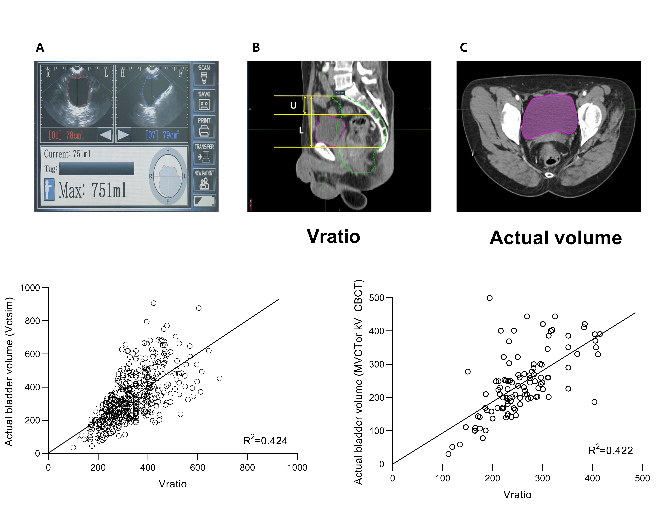
Background: Despite detailed instruction for full bladder, patients are unable to maintain consistent bladder filling during a 5-week pelvic radiation therapy (RT) course. We investigated the best bladder volume estimation procedure for verifying consistent bladder volume.Methods: We reviewed 462 patients who underwent pelvic RT. Biofeedback using a bladder scanner was conducted before simulation and during treatment. Exact bladder volume was calculated by bladder inner wall contour based on CT images (Vctsim). Bladder volume was estimated either by bladder scanner (Vscan) or anatomical features from the presacral promontory to the bladder base and dome in the sagittal plane of CT (Vratio). The feasibility of Vratio was validated using daily megavoltage or kV cone-beam CT before treatment.
Results: Mean Vctsim was 335.6 ± 147.5 cc. Despite a positive correlation between Vctsim and Vscan (R2 = 0.278) and between Vctsim and Vratio (R2 = 0.424), Vratio yielded more consistent results than Vscan, with a mean percentage error of 26.3 (SD 19.6, p < 0.001). The correlation between Vratio and Vctsim was stronger than that between Vscan and Vctsim (Z-score: - 7.782, p < 0.001). An accuracy of Vratio was consistent in megavoltage or kV cone-beam CT during treatment. In a representative case, we can dichotomize for clinical scenarios with or without bowel displacement, using a ratio of 0.8 resulting in significant changes in bowel volume exposed to low radiation doses.
Conclusions: Bladder volume estimation using personalized anatomical features based on pre-treatment verification CT images was useful and more accurate than physician-dependent bladder scanners.
Affiliations
Nalee Kim 1 2 , Hong In Yoon 1 , Jin Sung Kim 1 , Woong Sub Koom 1 , Jee Suk Chang 3 , Yoonsun Chung 4
1 Department of Radiation Oncology, Yonsei Cancer Center, Yonsei University Health System, Yonsei University College of Medicine, 50-1 Yonsei-ro, Seodaemun-gu, Seoul, 03722, Republic of Korea.
2 Department of Radiation Oncology, Samsung Medical Center, Sungkyunkwan University School of Medicine, 81 Irwon-ro, Gangnam-gu, Seoul, 06351, Republic of Korea.
3 Department of Radiation Oncology, Yonsei Cancer Center, Yonsei University Health System, Yonsei University College of Medicine, 50-1 Yonsei-ro, Seodaemun-gu, Seoul, 03722, Republic of Korea. changjeesuk@yuhs.ac.
4 Department of Nuclear Engineering, Hanyang University, 222 Wangsimni-ro, Seongdong-gu, Seoul, 04763, Republic of Korea. ychung@hanyang.ac.kr.
- 키워드
- Bladder volume; Computed tomography simulation; Cone-beam CT; Megavoltage CT.
- 연구소개
- 직장암 환자에서 5주간의 방사선 치료기간 동안 일정량의 방광 용적을 채우도록 교육하고, 초음파로 확인을 하지만, 그 정확성이 다소 부족한 것이 사실입니다. 저희 연구 논문에서는 설계용 CT 전 그리고 각 치료 전 MV 및 kv cone-beam CT를 바탕으로 개별 환자의 bladder dome과 base에 따른 해부학적 구조를 이용하여 방광 용적을 구하는 방법을 고안하였습니다. 해부학적 구조 비율로 구한 방광 용적은 초음파로 구한 용적보다 실제 용적에 더 가깝게 예측할 수 있음을 확인하였습니다. 그리고 그 비율에 따라 정상 장기의 선량 분포에도 차이를 보이는 것을 검증하였습니다. 시술자에 따라 다양한 결과를 보일 수 있는 다소 부정확한 초음파에 비하여 각 환자의 해부학적 구조의 비율을 바탕으로 한 방광 용적 예측의 가능성을 살펴보았기 때문에, 좀 더 정확하고 개별화된 방사선 치료를 하는 데 도움이 될만한 정보라고 생각합니다.시술자에 따라 다양한 결과를 보일 수 있는 다소 부정확한 초음파에 비하여 각 환자의 해부학적 구조의 비율을 바탕으로 한 방광 용적 예측의 가능성을 살펴보았기 때문에, 좀 더 정확하고 개별화된 방사선 치료를 하는 데 도움이 될만한 정보라고 생각합니다.
- 덧글달기
- 이전글 [Cancer Res Treat.] Intensity-Modulated Radiotherapy-Based Reirradiation for Head and Neck Cancer: A Multi-institutional Study by Korean Radiation Oncology Group (KROG 1707)
- 다음글 [Radiat Oncol.] Phase I/IIa trial of androgen deprivation therapy, external beam radiotherapy, and stereotactic body radiotherapy boost for high-risk prostate cancer (ADEBAR)


